
Superstars of science
The loud humming sound of machines at work pours out as group leader Péter Lénárt pulls open the two solid metal doors. We step into a dark anteroom, and he instructs me to slip disposable plastic booties that look like blue shower caps over my shoes. “Now we are very professional,” he says jokingly as we walk into the main room. “During my PhD this was a small system in the corner of the lab – look at it now!”
The source of the noise is revealed to be not computer servers nor fume hoods but dozens of water tanks, filled with aquatic creatures that seem a bit out of place in the hills near Heidelberg, more than 400 km from the sea. Bright red starfish the size of my face adorn the ocean-blue walls of one tank; another holds spindly, ivory-coloured starfish with spines all over their arms; a third contains small, dark green ones with striking orange spots along their backs. Across the room are shallow, trough-like containers each holding more than a dozen bulbous African clawed frogs that periodically clamber over each other. A pale purple sea urchin clings to the glass of its enclosure, tiny tube feet holding it in place. The room resembles an aquarium or a pet store rather than a science lab.

The right mix
Welcome to the marine facility of EMBL’s Laboratory Animal Resources department. True to Lénárt’s word, it is professional-grade: animal tanks are supplied with a 50/50 mixture of water from two 3000-litre containers that look like beer stills. One is filled with natural seawater that’s shipped in from the North Sea every month, the other with artificial seawater that contains a defined mixture of salts to maintain crucial parameters such as salinity and pH. The facility is equipped with all the pumps, filters, lights and other elements required to properly care for its residents. Staff members monitor the animals throughout the day, and ensure each tank is kept at the right temperature and salinity.
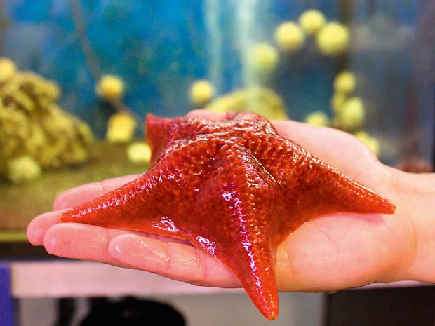
The big red ones are called bat stars… We ship them here by FedEx.
The marine facility is the brainchild of Detlev Arendt, a senior scientist at EMBL Heidelberg who studies neural development and evolution in the marine worm Platynereis dumerilii. In 2003, frustrated with the limitations of keeping specimens in buckets of seawater in the lab, he spearheaded the construction of the current system, which provides everything needed to breed and maintain large numbers of his model organism in a controlled, consistent environment. “Our lab sort of jumped onto that project and connected our system to his,” says Lénárt, gesturing to the starfish tanks. “The big red ones are called bat stars, and they are quite common along the west coast of the United States. We ship them here by FedEx.” The green and orange starfish are similarly well travelled: Lénárt had these sent from Japan rather than the States, due to concerns over a recent decline in the starfish population in California that could threaten the health of the lab’s supply.
Stars of the show
Lénárt’s lab studies meiosis, the process by which a reproductive cell divides into gametes – spermatocytes in males or oocytes in females – that can then combine during fertilisation. Whereas meiosis in male starfish results in four equally sized spermatocytes, the female lineage produces one very large egg cell that is packed with nutrients to support a developing embryo. “One of the main things we are interested in is how division happens in these exceptionally large cells,” says Lénárt. “It’s extremely asymmetrical.”
With most of the cellular contents concentrated in one cell, the remaining three meiosis products become small ‘polar bodies’ whose only purpose is to serve as reservoirs for the extra DNA that needs to be jettisoned from the oocyte. “From an engineering point of view, it’s like a scaling problem: how do you scale up cell division from a small cell to a large cell, and with such asymmetry?” Lénárt says.
Meiosis is a difficult process to control and image in the lab, particularly in mammals. Mice, one of the most widely used model organisms, present many challenges: they must be ordered weeks in advance so that they are the right age to produce oocytes; the oocytes must be removed from the mouse’s body and kept at 37˚C during their transformation into an egg cell; and the process takes about eight hours to complete, meaning that the imaging microscope must somehow be automated to keep track of the oocytes. Starfish, by contrast, are much easier to work with. They are one of the few animals whose maturation-inducing hormone has been identified, which allows researchers to control when the oocytes start to mature into eggs. “We just keep them at room temperature in sea water, we add the hormone, and the whole process takes one and a half hours. It’s a system which allows you to do ‘discovery’ research and explore new things, because you can proceed quickly,” says Lénárt.

Intriguingly, Lénárt and his team are discovering a large amount of diversity among oocytes of different species. “What is really exciting to me is that oocytes are single-celled in all animals,” he says. “Normally adaptation to the environment happens at the organism level. But in the egg, it happens at the single-cell level.” Different organisms’ eggs are subjected to widely varying conditions: some float in the sea, some lay buried in desert sand, and some are contained within the mother’s body until birth. Comparing different animals’ cells at the same early developmental stage can offer insights into how they evolved into the fantastically varied pantheon of life that exists on the planet today.
Unexpected discoveries
Specifically, research on starfish oocytes has revealed diversity at the level of core cellular machineries that are highly conserved across very different organisms. “Now we are looking at how different cell types combine those common elements to achieve different cellular functions. It’s like using the same set of Lego blocks to build either a castle or a pirate ship,” says Lénárt. His group has found, for example, that an actin network used for cell migration in somatic cells has a different role in starfish oocytes: it’s needed to break the nuclear envelope of the exceptionally large oocyte nucleus during cell division. This and other unexpected observations are what keep Lénárt and his group on their toes.

Starfish are rarely used to do this type of investigative research, which is often conducted in yeast or fruit fly cells. Lénárt is part of a growing community of scientists who are looking beyond established ‘model organisms’ and focusing their research on other creatures to explore nature’s diversity. Detlev Arendt’s lab was the first at EMBL to use Platynereis as its primary animal model, and has recently expanded its scope to include animals such as amphioxus lancelet fish and the starlet sea anemone Nematostella vectensis. These creatures are attractive for scientific study, not only because of their evolutionary significance – all of them have relatively simple neural networks – but because they lend themselves well to being grown and studied in the lab. “When you choose a new model organism, you have to consider not only the biological questions, but all the technical things as well,” says Marzia Sidri, a project manager in Arendt’s lab. “In general, it is much more difficult to work with animals that grow too slowly or aren’t easy to image.”
Beneath the surface
Across the Heidelberg campus in the EMBL Advanced Training Centre, a computer monitor flickers to life in the training lab, displaying what look like tiny glass satellites floating above a black background. Switching between looking at the screen and through a microscope, Ina Arnone deftly manoeuvers two micropipetting needles into view using a series of six knobs and dials that she manipulates with practised ease. Three students in white lab coats peer over her shoulder, watching their instructor’s every move. The needles converge on one of the delicate shapes and attempt to pin it in place. The first needle bumps the target so that it spins away in infuriatingly lazy circles, forcing Arnone to start all over again. She patiently begins readjusting the needles, one knob at a time, her calm determination evidence of her expertise. Finally, she moves one of the needles close enough to gently suction the satellite to it, which she does by sucking on a thin rubber tube attached to the needle. While that needle holds its prey in place, Arnone slowly moves the other one forward. The glassy shape resists slightly, and then the needle finally pokes its way through. The students let out an audible sigh of relief, while Arnone sits back from the microscope looking as relaxed as if she’d just sliced a tomato for lunch. “And that’s how you microinject a sea urchin larva,” she says.
And that’s how you microinject a sea urchin larva
Arnone has her own research group at the Stazione Zoologica Anton Dohrn in Naples, Italy, where she’s investigating the evolutionary and developmental biology (evo-devo) of gene expression and neurology. Her primary model organism is the sea urchin, principally Strongylocentrotus purpuratus, the entire genome of which was sequenced in 2006, and Paracentrotus lividus, which is abundant in the waters near Naples. She has brought her purple, spiky specimens to EMBL Heidelberg for a week-long training course sponsored by the Neptune Consortium, a network created by the Marie Skłodowska-Curie Actions fund to train young scientists who are studying evo-devo in marine organisms. The fact that marine animals’ eggs and larvae are easy to produce, largely transparent and develop relatively quickly has led many scientists like Arnone to look to the sea when choosing new model organisms to study. Her other Neptune partners have brought their own alternative model organisms as well, including the common jellyfish Clytia hemisphaerica and Arendt’s Platynereis worms, turning the training lab into a temporary menagerie.

Students on the course practise injecting living, developing embryos with various substances that alter which genes are expressed, either by inserting new DNA or by blocking or enhancing specific genetic products (RNAs and proteins). Looking at how these modified embryos develop compared to normal embryos can help researchers understand the function of a given gene or pathway. Investigating these processes in primitive animals like sea urchins and jellyfish can also provide insight into how early body plans gave rise to the more complex features of ‘higher’ animals.

“Some changes that accrue in the embryo as it develops reflect the changes that have accumulated on the evolutionary scale,” says Sidri, who helped organise the Neptune course. The embryos of many land animals, like chickens and humans, have primordial gill slits and neck arches that they lose as they develop, just as the ancestors of birds and mammals lost theirs when they evolved from fish-like ancestors millions of years ago. “That’s the basis of the whole evo-devo field,” says Sidri. Although evo-devo model techniques are commonly used on well-established model organisms such as Drosophila, testing them on different species can reveal information that fruit flies might not be particularly well-suited to provide. “The reason why scientists are looking for new models is that you can’t generalise the structure and function of all animals just by analysing one species. You need to analyse more animals so you can compare and understand more about them all,” says Sidri.
Living organisms aren’t the only source of insight: Giannis Kesidis, one of the students attending the Neptune course, is a paleontologist at Uppsala University in Sweden who studies arthropod fossils from the Cambrian period. Although his work doesn’t directly involve microinjecting larvae, “it’s very useful to know what the developmental biologists are working on, because their hypotheses often have evolutionary implications that lead them to collaborate with people like me, who work on the fossil record,” he says.
Changing tides
Back in the marine facility, Lénárt shows me its newest addition: tiny jellyfish polyps donated by Evelyn Houliston, a researcher based at the Villefranche Oceanographic Laboratory in France who attended the Neptune course that week. Scarcely larger than a grain of rice, they’re affixed to a clear plastic slide submerged in a small container of seawater hidden away in a makeshift cupboard. “We’re still working on setting this up, so it’s under construction,” Lénárt says with a tinge of embarrassment, as if apologising for the polyps’ juvenile state.
Establishing a new animal line for study takes some time, but Lénárt is excited about the insights another organism could offer for his research. “The jellyfish is very close to the bottom of the animal evolutionary tree, so we are hoping to see some of the very basic features of animal oocytes by studying it. Also, the polyp form reproduces by budding and grows on a glass plate just like plants, which offers great opportunities for genetic manipulation,” he says.

Lénárt has watched the evo-devo field branch out to include animals beyond those typically used as model organisms over the past few years. “I think cell biology has been largely focusing on conserved features, and it is often argued that when we find a conserved mechanism in yeast or Drosophila or mice, we can apply whatever we learn about it directly to human cells,” he says. “We are now at a stage where people are taking interest in the diversity of those mechanisms themselves.”
Lénárt gazes at the squat, orange-spotted Japanese starfish sitting next to the large, spiky-armed ones. “While what happens in a starfish oocyte might not be exactly the same as what happens in a human egg, we are learning a lot about core cellular machineries by looking at their different functions in different places,” he says. “It’s a great way to learn about how those cellular mechanisms work which, at a basic level, seems to be pretty universal across cells in all species of life on Earth.”


