17 July 2026

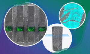
Researchers have designed synthetic and inducible proteins that bind to bright fluorescent dyes with high specificity and affinity, significantly expanding the toolkit for multicolour imaging of proteins inside cells.
SCIENCE & TECHNOLOGY
17 February 2025
New funding from the Chan Zuckerberg Initiative (CZI) supports two multidisciplinary projects across EMBL’s units and sites to support the development of imaging technologies.
SCIENCE & TECHNOLOGY
16 December 2024
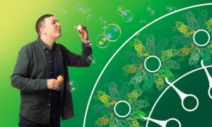
Veijo Salo, postdoc at EMBL Heidelberg, talks about seipin, the cell’s molecular ‘bubble blower’.
PEOPLE & PERSPECTIVES
26 October 2023

Jan Kosinski, Julia Mahamid, and Georg Zeller have received grants to enable ambitious projects aimed at mapping the cellular protein synthesis machinery in context and understanding complex host-microbiome interactions, respectively.
EMBL ANNOUNCEMENTS
2023
embl-announcementsscience
4 July 2023

Three EMBL group leaders and six EMBL alumni were recognised for their contributions to the life sciences.
LAB MATTERSPEOPLE & PERSPECTIVES
2023
lab-matterspeople-perspectives
2 February 2023
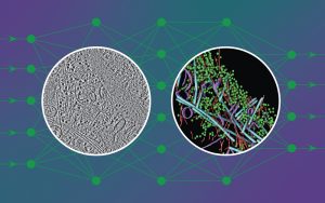
New artificial intelligence tool adds speed and detailed cellular information to analysis of cryo-electron tomography to aid researchers’ understanding of inner cell workings.
28 September 2022
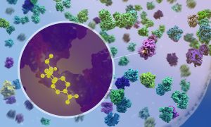
New research by EMBL scientists shows at atomic detail how antibiotics affect the process of protein production inside bacteria.
SCIENCE & TECHNOLOGY
2022
sciencescience-technology
21 December 2021
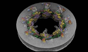
EMBL Hamburg’s Kosinski Group, the Beck Laboratory at the Max Planck Institute of Biophysics, and colleagues at EMBL Heidelberg recorded the nuclear pore complex contracting in living cells. They visualised the movement with an unprecedented level of detail with help of new software called…
SCIENCE & TECHNOLOGY
2021
sciencescience-technology
23 February 2021
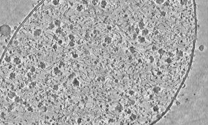
Liang Xue used cryo-electron tomography to capture this detailed image of a Mycoplasma pneumoniae cell.
SCIENCE & TECHNOLOGY
2021
picture-of-the-weekscience-technology
2 February 2021
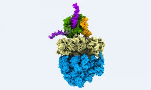
This colourful image shows biological information flow in action: It’s a supramolecular assembly of DNA, RNA and proteins, observed directly inside a bacterial cell while turning genetic information into protein.
SCIENCE & TECHNOLOGY
2021
picture-of-the-weekscience-technology
7 December 2020
While cryo-electron tomography (cryo-ET) was first envisioned in 1968, the advances the Mahamid group are bringing to this 3D method for studying molecules directly inside cells are new, and are likely to greatly expand its use.
SCIENCE & TECHNOLOGY
2020
sciencescience-technology
No results found