New software helps researchers decode the cellular architecture of lymphoma
An open-source analysis framework developed by researchers at EMBL, HHU, and other institutions makes it easier to study how cells are organised within tumours, paving the way for new insights into cancer biology
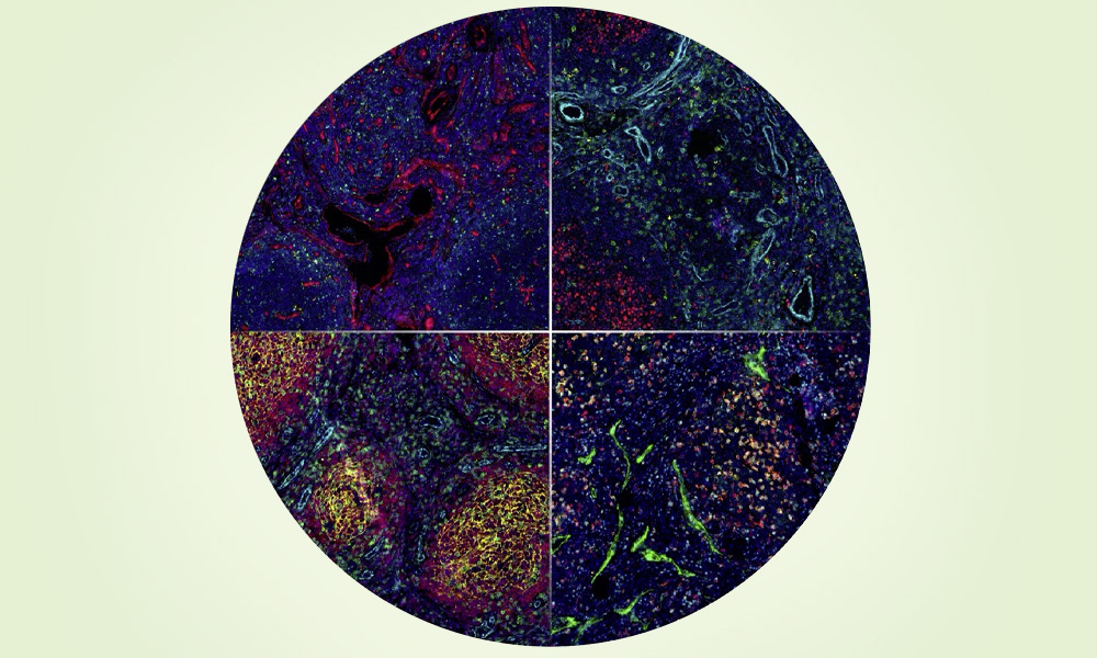
Multiplexed imaging captures numerous proteins simultaneously in a single tissue sample, such as a lymphoma biopsy, and makes different cell types and tissue structures visible. These can then be analysed systematically and reproducibly using the spatialproteomics software. Credit: UKD
Summary
EMBL and collaborators developed ‘spatialproteomics’, an open-source software that helps researchers analyse complex tissue images and understand how cells are organised within tumours.
The tool streamlines a workflow that was previously fragmented, taking users from raw microscopy images through cell segmentation, annotation, and spatial analysis, to biological insight in one unified framework.
In studies of B-cell non-Hodgkin lymphoma, the software revealed distinct spatial patterns in tumour and immune cells that helped distinguish between slow-growing and aggressive samples.
Spatialproteomics could be useful in studies across many cancers, inflammatory diseases, and in tissues where spatial organisation matters.
How cancer develops and responds to treatment depends not only on the tumour cells themselves but also on the complex ecosystem of surrounding immune cells, blood vessels, and connective tissue. In B-cell non-Hodgkin lymphoma, these cellular neighbourhoods can determine whether a tumour grows, evades the immune system, or responds to therapy. Understanding this spatial organisation has become one of the major challenges in cancer research.
Now, researchers from the European Molecular Biology Laboratory (EMBL), Heinrich Heine University Düsseldorf (HHU), and University Hospital Düsseldorf (UKD) have developed spatialproteomics, an open-source software package that makes it much easier to analyse highly complex tissue images. Published in Nature Methods, the software enables researchers to move seamlessly from microscopy images to biological insights, helping reveal how different cell types are organised within healthy and diseased tissues.
“Modern imaging technologies allow us to observe tissues in extraordinary detail,” said Matthias Meyer-Bender, first author of the study and PhD student at EMBL. “But turning these images into biological knowledge has remained technically challenging. We developed spatialproteomics to make these analyses more accessible, reproducible, and scalable.”
Recent advances in multiplexed imaging allow researchers to map thousands to millions of individual cells within a single tissue sample. Unlike conventional pathology methods, which typically examine only a handful of cellular features, these technologies simultaneously capture dozens of molecular markers while preserving the spatial arrangement of cells. This provides an unprecedented view of the tumour microenvironment and the interactions that shape disease.
However, analysing these datasets requires identifying, classifying, and quantifying vast numbers of cells before researchers can investigate their spatial arrangement. Until now, this process often involved combining multiple software packages and custom-built analysis pipelines.
Spatialproteomics brings these steps together into a single workflow. The software guides researchers from raw microscopy images through cell segmentation, annotation, and spatial analysis, making sophisticated image analysis more efficient and easier to reproduce.
To demonstrate the framework’s capabilities, the team analysed tissue samples from patients with B-cell non-Hodgkin lymphoma. Spatialproteomics enabled the researchers to characterise the spatial distribution of proteins and identify distinct patterns in the organisation of tumour and immune cells. These spatial signatures supported the distinction between indolent (slow-growing) and aggressive lymphomas, illustrating how computational analysis can help uncover biologically and clinically relevant tissue features.
“Accessible computational tools are essential if advanced imaging technologies are to realise their full potential,” Meyer-Bender said. “By lowering technical barriers and standardising analysis workflows, spatialproteomics allows more researchers to extract meaningful information from complex imaging data.”
Although demonstrated using lymphoma samples in this study, the researchers expect the software to benefit many areas of biomedical research.
“Spatialproteomics is not limited to lymphoma research,” said co-senior author Peter-Martin Bruch of Heinrich Heine University Düsseldorf and University Hospital Düsseldorf. “The framework can be applied to many types of cancer as well as inflammatory diseases and other complex tissues where spatial organisation plays a critical role.”
The project brought together complementary expertise across institutions. Clinical and experimental researchers in Düsseldorf collaborated closely with EMBL scientists specialising in bioinformatics, data science, and software engineering to develop a tool that addresses both methodological challenges and practical biomedical questions.
“This project demonstrates the value of interdisciplinary collaboration,” Bruch said. “By combining clinical expertise with EMBL’s strengths in computational biology and software development, we created a framework that is both technically robust and directly relevant to biomedical research.”
Spatialproteomics is freely available as open-source software, allowing researchers worldwide to integrate it into their own imaging workflows and further expand its capabilities. In addition to EMBL, HHU, and UKD, researchers from Heidelberg University and the Center for Integrated Oncology (CIO) Aachen Bonn Cologne Düsseldorf contributed to the study.