New services in 2026 – Pilot projects wanted!
Following the full implementation of ‘Serial lift-out for cryo-electron tomography’, the next two services we plan to add to our portfolio are Oblique Plane Microscopy (OPM), and Brillouin Microscopy, both of which are aimed at enabling investigations in living cells and organisms. The microscopes are custom-built based on developments in academic imaging technology development labs (further details below). Covering fast dynamics in living systems, and the mechanical properties of tissues, these new capabilities enable researchers to explore biology in its full dynamic, and physical complexity. Combined with ‘serial lift-out’ those discoveries could be directly connected to structural understanding from the same cells or organisms.
We warmly invite all researchers whose scientific questions could benefit from Oblique Plane Microscopy and Brillouin Microscopy to contact us at ic-contact@embl.de and explore whether this could become a pilot project!
Oblique Plane Microscopy (OPM)
Fast, gentle volumetric imaging of living systems
Oblique Plane Microscopy (OPM) is a light-sheet fluorescence microscopy technique that enables rapid, gentle three-dimensional imaging of living samples. Unlike conventional light-sheet systems, OPM uses a single objective lens to both illuminate an obliquely oriented light sheet and collect emitted fluorescence, allowing imaging in an inverted configuration and with conventional sample mounting from the top [1,2].
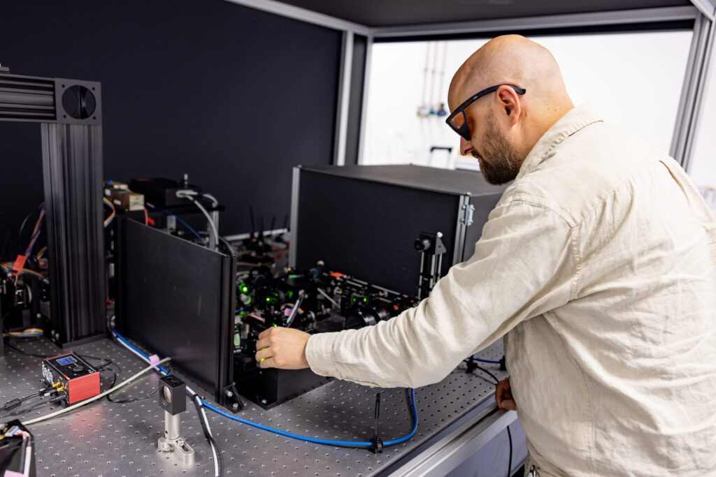
Rory Power, Senior Engineer: “Uniquely, OPM enables high-numerical-aperture and ultra-fast volume imaging in coverslip-mounted samples, though this comes with challenges for stability and day-to-day operability. The system I built at the EMBL Imaging Centre overcomes these challenges by combining ultra-stable microscope rooms, precision mechanical design and fabrication, and innovations in focus control, resulting in a system that requires minimal adjustment and supports routine operation.”
This makes OPM ideally suited for users who want the advantages of light-sheet imaging — speed, intrinsic 3D imaging capabilities and low photobleaching/phototoxicity — without complex sample preparation. The IC’s implementation is optimised for high-numerical aperture for high spatial resolution and sensitivity, making it particularly powerful for observing fast subcellular dynamics in thin 3D samples up to ~30 µm thick.
Brillouin Microscopy
Label-free imaging of mechanical properties
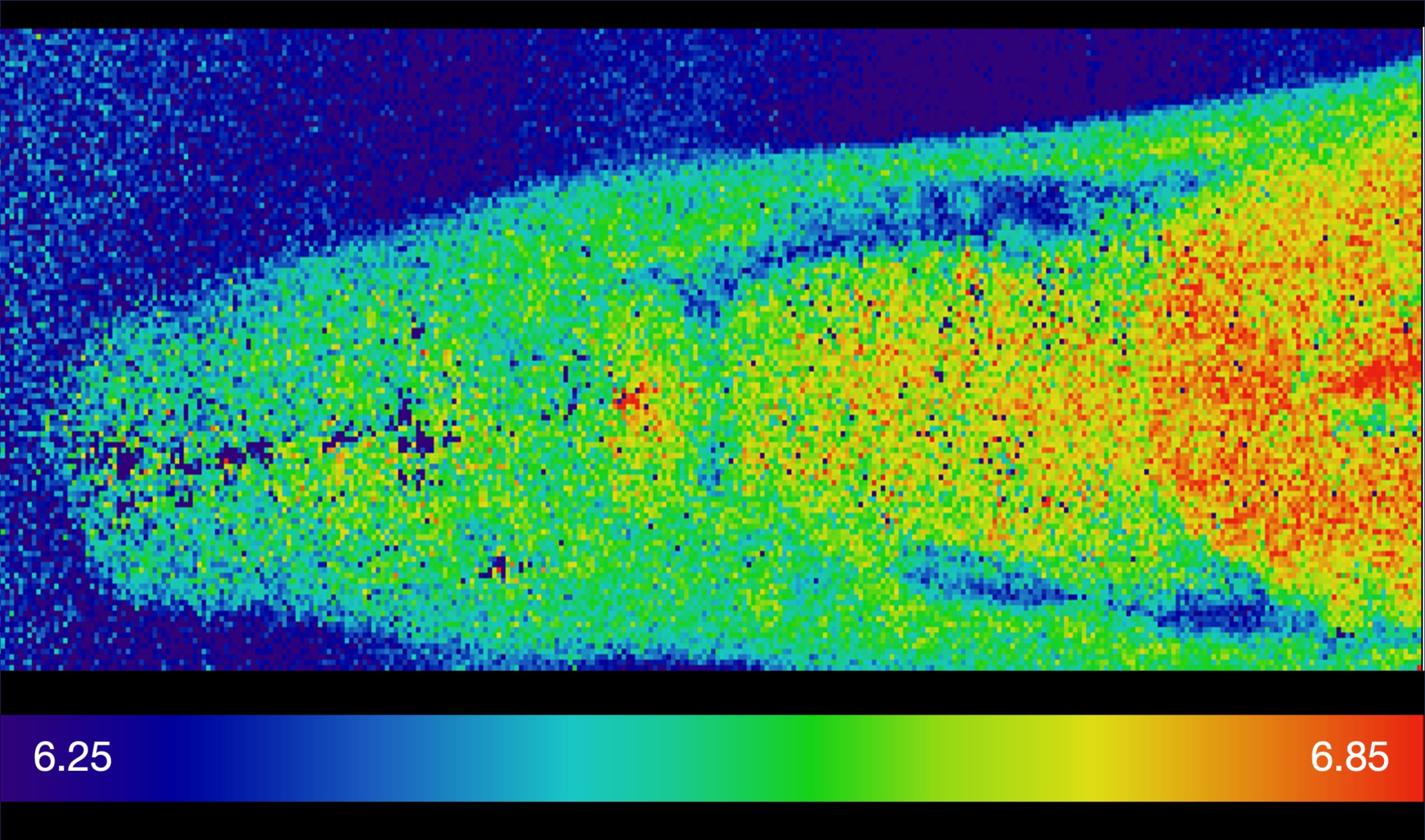
Brillouin microscopy is a non-invasive, label-free technique that measures the mechanical properties of biological samples, such as stiffness and viscosity. It is based on Brillouin scattering, a subtle frequency shift that occurs when light interacts with acoustic vibrations inside a material [3,4,5,6].
Our Brillouin microscope builds on developments from Robert Prevedel’s group at EMBL Heidelberg and is fully integrated with a high-end confocal fluorescence microscope, enabling multimodal imaging of structure, composition, and mechanics within the same sample.
This approach opens new possibilities for studying mechanobiology, tissue organisation, and disease-related changes in material properties, without the need for external probes or labels.
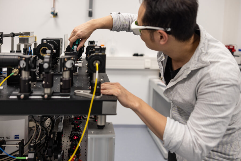
Tzu-Lun Wang, Engineer: “The Brillouin microscope at the EMBL Imaging Centre is integrated with a high-end confocal fluorescence microscope. This combination enables users to investigate the mechanical properties of biomaterials within a high-definition spatial map highlighted by molecular markers in tissues.”
Enabling discovery through advanced imaging!
With the implementation of ‘Serial lift-out’, OPM and Brillouin microscopy we keep on pushing our mission forward—making advanced imaging tools and expert support more accessible to the life-science community. Spanning molecular assemblies within cells, fast dynamics in living systems, and the mechanical properties of tissues, these new capabilities enable researchers to explore biology in its full structural, dynamic, and physical complexity.
References
[1] https://doi.org/10.1364/OE.16.020306
[2] https://doi.org/10.7554/eLife.57681
[3] https://doi.org/10.1364/boe.10.001420
[4] https://www.nature.com/articles/s42003-021-02662-5