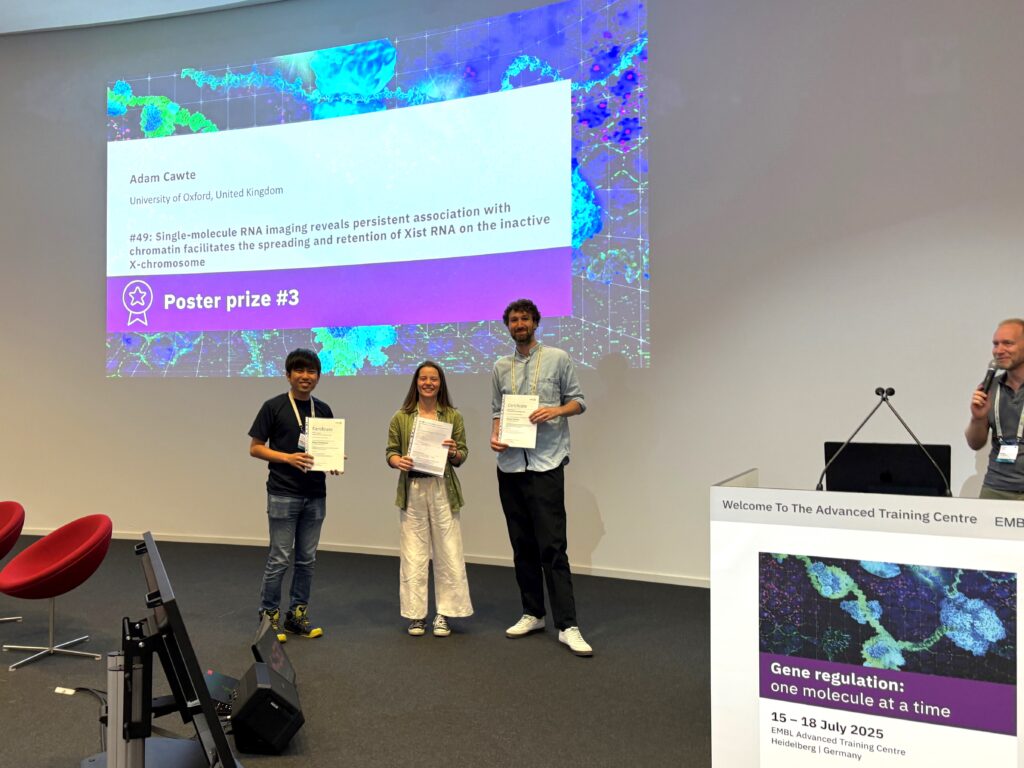
Best poster prizes at ‘Gene regulation: one molecule at a time’
The EMBL Conference ‘Gene regulation: one molecule at a time‘ took place in July at EMBL Heidelberg and virtually.
The last few years have seen the independent emergence of microscopy and genomics technologies to monitor gene regulation processes with single molecule resolution. Recording the dynamic function of regulatory proteins on DNA and RNA now opens opportunities to understand key parameters of the process, such as kinetics, or how various activities are combined in vivo.
Our brand-new conference brought together pioneers of the single molecule microscopy and single molecule genomics fields, as well as theoretical biologists, to catalyse the emergence of a new generation of mechanistic models of gene regulation across each step of the central dogma.
For the inaugural edition of the meeting, we had 100 people attending on-site and 43 participants attending remotely. There were 10 fellowships provided by the EMBL Corporate Partnership Programme. With the total of 61 posters to view, we held two poster sessions during which the presenters could discuss their research — their work was then voted for by all participants. There were three poster prizes awarded during the meeting. We are pleased to share with you two of the winners’ abstracts!
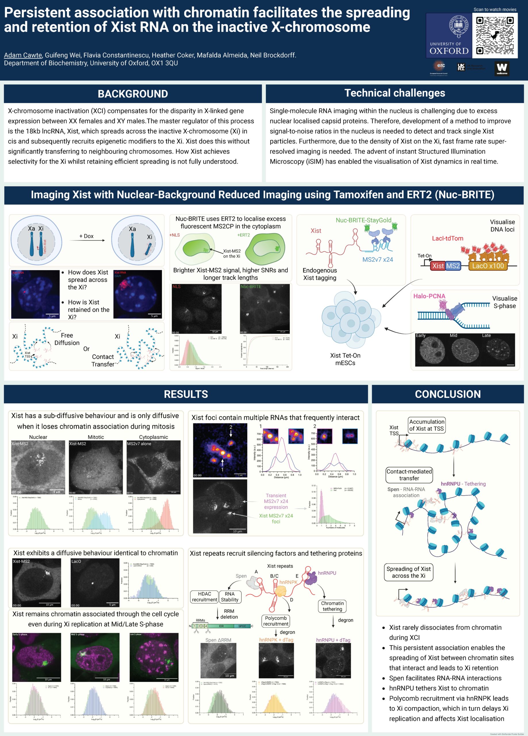
Single molecule RNA imaging reveals persistent association with chromatin facilitates the spreading and retention of Xist RNA on the inactive X chromosome
Presenter: Adam Cawte
Authors: Adam Cawte, Guifeng Wei, Heather Coker, Mafalda Almeida, Flavia Constantinescu, Neil Brockdorff

University of Oxford, UK
Abstract:
X chromosome inactivation (XCI) is a mammalian dosage compensation mechanism that compensates for the disparity in X linked gene expression between XX females and XY males. During XCI in females, the majority of genes on one of the X chromosomes are epigenetically silenced. The master regulator of this process is the 18kb lncRNA, Xist, which spreads across the inactive X chromosome (Xi) in cis and subsequently recruits epigenetic modifiers to the Xi. Xist does this without significantly transferring to neighbouring chromosomes. How Xist achieves selectivity for the Xi whilst retaining efficient spreading is not fully understood.
Single molecule RNA imaging within the nucleus is challenging due to excess background fluorescence of nuclear localised MS2 capsid proteins. Therefore, we developed a method to significantly reduce this excess by using cytoplasmic retained capsid proteins. This improves signal to noise ratios and allows for the accurate detection and tracking of single Xist particles at fast frame rates with super resolved instant Structured Illumination Microscopy.
With this technique, we can dissect the different diffusive behaviours Xist RNA particles use when spreading across the Xi. We observe that the majority of Xist particles are chromatin associated, and their dynamics correlate well with the underlying movements of chromatin and DNA DNA contacts. We propose that Xist primarily spreads across the Xi via a series of “hand over” events between frequently interacting DNA loci. This model allows for Xist to spread efficiently during XCI whilst retaining selectivity for the Xi. Furthermore, we have investigated how key players involved in RNA tethering and stability affect Xist dynamics and spreading.
Linker histone H1 functions as a liquid like glue to organize chromatin in living human cells
Presenter: Masa Shimazoe
Authors: Masa Shimazoe, Satoru Ide, Sachiko Tamura, Kazuhiro Maeshima
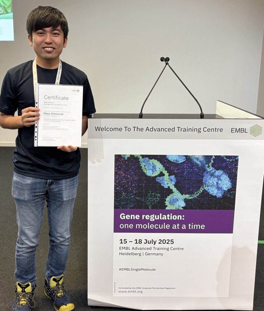
National Institute of Genetics, Japan
Abstract:
Linker histone H1 is the most abundant chromatin protein known to compact chromatin. How can H1 compact chromatin? A textbook model tells us that H1 compacts chromatin via stabilization of the 30-nm chromatin fiber, where nucleosomes are regularly folded like a solenoid or zigzag [1, 2]. However, studies over a decade show the regular 30-nm chromatin fiber is not the basic structure of chromatin in living cells. Nucleosomes are thought to be irregularly folded into clusters called chromatin domains [3]. Thus, we need to revise the textbook model on how H1 compacts chromatin in the dynamic chromatin domain. Here, to reveal how H1 works in chromatin compaction, we performed single-molecule imaging and tracking of H1 in living human cells. Single-molecule imaging demonstrated more dynamic H1 motion than nucleosomes, consistent with previous reports [4][5]. To further investigate H1 behavior, using a machine-learning-based analysis (vbSPT) [6], we classified H1 trajectories into three states: “bound,” “liquid-like,” and “dissociated,” which reflect static “dyad” H1 binding on a nucleosome, multivalent H1 interactions with several nucleosomes, and temporal dissociation from chromatin, respectively. This observation can explain both in vitro studies showing static H1 binding to a nucleosome and in vivo observation of dynamic H1 behavior. “Liquid-like” H1 makes up 60% of trajectories, suggesting a major contributor to H1 function.
Furthermore, by combining single H1 imaging and PALM imaging of nucleosomes, we found “liquid-like” H1 diffuses mainly within the chromatin domain while weakly interacting with several nucleosomes. Taken together, we conclude the majority of H1, which has “liquid-like” behavior via multivalent interactions, serves as liquid-like “glue” for the chromatin domain [7]. Such a liquid-like “glue” property of H1 allows the chromatin domain to be compacted and accessible simultaneously so that other molecules, including transcription factors, can penetrate the domain.
[1] J. T. Finch et al. PNAS (1976) [2] F. Song et al. Science (2014) [3] K. Maeshim et al. Curr. Opin. Cell. Biol. (2019) [4] T. Misteli et al. Nature (2000) [5] M. A. Lever et al. Nature (2000) [6] F. Persson, M. Linden et al. Nat Methods (2013) [7] M. A. Shimazoe et al, bioRxiv (2025)
Find out more about the #EMBLSingleMolecule conference from the blog post written by Priyanka Yadav, who participated as an event reporter!
The EMBL Conference ‘Gene regulation: one molecule at a time‘ took place from 15 – 18 July 2025 at EMBL Heidelberg and virtually.