Sync to grow
Oscillation of gene activity may underlie how embryos grow in proportion
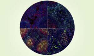
Gene expression wave in the lower part of the future vertebrae column of a mammalian embryo. As the wave goes forward, new pre-vertebrae are formed and the future vertebrae column elongates. (Image and video credit: Nature)
In a nutshell:
- The size of pre-vertebrae in a mammalian embryo is controlled by a wave-like gene expression pattern along its back
- The size of each pre-vertebra is proportional to the speed of the wave: the faster the wave, the bigger the vertebra
- Embryonic cells coordinate gene activities with each other to adapt the size of pre-vertebrae to the overall size of the embryo
From a single-cell egg to a fully functional body: as embryos develop and grow, they must form organs that are in proportion to the overall size of the embryo. The exact mechanism underlying this fundamental characteristic, called scaling, is still unclear. However, a team of researchers from EMBL Heidelberg is now one step closer to understanding it. They have discovered that scaling of the future vertebrae in a mouse embryo is controlled by how the expression of some specific genes oscillates, in a coordinated way, between neighbouring cells. Published today in Nature, their findings highlight how important this oscillatory pattern, and its regulation, is to ensure that embryos grow up to become well-proportioned animals.
Neighbouring cells in the future vertebral column of an embryo coordinate to turn specific genes on and off in turn, thus generating a wave of gene expression similar to the ‘Slide to unlock’ animation on your smart phone. To study this process, and determine its impact on how the relative sizes of the future vertebrae are maintained, the researchers developed a new technique.
“Using this new assay, we were able to film this wave of gene expression in real time with high precision, and to identify whether this pattern could change according to the overall size,” explains Alexander Aulehla who coordinated the study at EMBL Heidelberg. “There is a clear link: when the embryo is smaller, the number of segments formed remains the same, but each segment is smaller and the expression waves are proportionally slower.”
The speed of the wave seems to be the essential characteristic to predict the size of the future vertebra: the faster the wave, the bigger the vertebra. Similar expression waves have been observed in several vertebrates and also in insect species, so this communication pattern amongst embryonic cells seems to be very wide-spread. However, scientists haven’t yet elucidated how the speed of the wave is controlled at a molecular level.
The technique developed in this study might be the key to helping the team understand this complex and fundamental mechanism. In order to make observation easier, the scientists grew only one layer of embryonic stem cells to which a specific marker was added, to follow the expression of the Notch genes. The combination of the monolayer and marking made real-time observation of gene expression possible. In the future this new technique might help researchers understand the details of how embryonic cells sync to grow.
The video is also available on the EMBL YouTube Channel.
Additional resources
Listen to the Nature podcast that features this finding (min: 9:33)
Read the release distributed by McGill University
More information on the research of the Aulehla Group
Source article
Scaling of embryonic patterning based on phase-gradient encoding – Volker M. Lauschke, Charisios D. Tsiairis, Paul François & Alexander Aulehla – Published online in Nature on 19 December, 2012.
Article abstract
A fundamental feature of embryonic patterning is the ability to scale andmaintain stable proportions despite changes in overall size, for instance during growth. A notable example occurs during vertebrate segment formation: after experimental reduction of embryo size, segments form proportionally smaller, and consequently, a normal number of segments is formed. Despite decades of experimental and theoretical work, the underlying mechanism remains unknown. More recently, ultradian oscillations in gene activity have been linked to the temporal control of segmentation; however, their implication in scaling remains elusive. Here we show that scaling of gene oscillation dynamics underlies segment scaling. To this end, we develop a new experimental model, an ex vivo primary cell culture assay that recapitulates mouse mesoderm patterning and segment scaling, in a quasi-monolayer of presomitic mesoderm cells (hereafter termed monolayer PSM or mPSM). Combined with real-time imaging of gene activity, this enabled us to quantify the gradual shift in the oscillation phase and thus determine the resulting phase gradient across the mPSM. Crucially, we show that this phase gradient scales by maintaining a fixed amplitude across mPSM of different lengths. We identify the slope of this phase gradient as a single predictive parameter for segment size, which functions in a size- and temperature-independent manner, revealing a hitherto unrecognized mechanism for scaling. Notably, in contrast to molecular gradients, a phase gradient describes the distribution of a dynamical cellular state. Thus, our phase-gradient scaling findings reveal a new level of dynamic information-processing, and provide evidence for the concept of phase-gradient encoding during embryonic patterning and scaling.