Edit

Tag:

picture release

Year
13 September 2009 In normal skin (left), the stem cells at the base, shown in green, differentiate into skin cells, shown in red. In mice whose skin has neither C/EBPα nor C/EBPβ (middle), this differentiation is blocked: green-labeled stem cells appear in upper layers of skin, and there are no differentiated skin cells (no red staining). This also happens at the initial stages of basal cell carcinomas. In skin where C/EBPα is present but has lost its capacity to interact with E2F, a molecule that regulates the cell cycle (right), skin cells start differentiating abnormally, before they have properly exited the stem cell ‘program’ (yellow/orange). This is similar to what is observed in the initial stages of squamous cell carcinomas, a more aggressive and invasive skin tumour.

How stem cells make skin

Science 
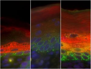
Stem cells have a unique ability: when they divide, they can either give rise to more stem cells, or to a variety of specialised cell types. In both mice and humans, a layer of cells at the base of the skin contains stem cells that can develop into the specialised cells in the layers above.…

2009

science

25 June 2009 The microscope image of the dorsal closure of a fly embryo shows alternating stripes of epithelial cells with aligned microtubule bundles (green) and epithelial cells treated with a microtubule-destroying drug (blue). Labelled in red is the protein actin that lines the border of cells, particularly the amnioserosa cells occupying the eye-shaped opening.

Uncovering how cells cover gaps

Science 
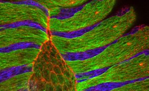
Researchers at the European Molecular Biology Laboratory (EMBL) in Heidelberg, Germany, came a step closer to understanding how cells close gaps not only during embryonic development but also during wound healing. Their study, published this week in the journal Cell, uncovers a fundamental…

2009

science

23 June 2009 Lattice maps for immature HIV particles. The 3D computer reconstruction shows the immature Gag lattice of HIV that matures to form the protein shell of the infecious virus. Maps are shown in perspective such that hexamers on the rear surface of the particle appear smaller. The side of the particle toward the viewer lacks ordered Gag. IMAGE: John Briggs/EMBL

New electron microscopy images reveal the assembly of HIV

Science 
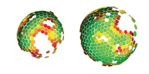
Scientists at the European Molecular Biology Laboratory (EMBL) and the University Clinic Heidelberg, Germany, have produced a three-dimensional reconstruction of HIV (Human Immunodeficiency Virus), which shows the structure of the immature form of the virus at unprecedented detail. Immature HIV is…

2009

science

24 February 2009 A full body shot of Medaka juveniles, taken by Philipp Keller, from the lab of Ernst Stelzer at the European Molecular Biology Laboratory (EMBL), with a newly developed microscope called Digital Scanned Laser Light Sheet Fluorescence Microscope. Picture credits: Philipp Keller, Stelzer Group, EMBL

Picture Release

Science 
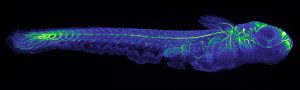
‘Useless fish with big eyes’. This is what Medaka, the name of the Japanese killifish in the pictures, means in Japan where it originally comes from. While its eyes are undeniably big, the fish has proven remarkably useful for scientists. It is a simple model organism, amenable to…

2009

science

No matching posts found

EMBLetc.

Looking for past print editions of EMBLetc.? Browse our archive, going back 20 years.

EMBLetc. archive

Newsletter archive

Read past editions of our e-newsletter

For press

Contact the Press Office
Edit