
New technique for imaging the life aquatic
New method for 3D imaging microorganisms lends insight into the creatures that inhabit our oceans
Many of nature’s diverse microorganisms have never actually been seen by researchers. That includes the miniscule predators, photosynthesisers, and symbionts that call the ocean home. A new automated microscopy technique makes it faster to 3D image and correctly classify microorganisms in marine samples. EMBL’s Luis Pedro Coelho explains the new technique and how this could lead to a better understanding of how some of the ocean’s smallest creatures live.
What did you do?

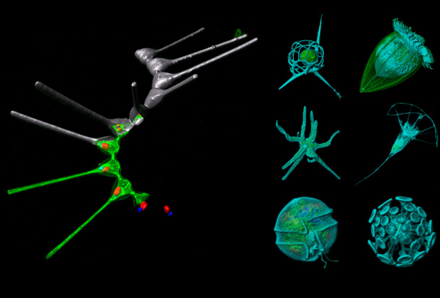
We developed a quick method to get detailed 3D images of diverse organisms in any sample taken directly from the environment. In our study, we looked at microscopic eukaryotes, a type of nucleus-containing organism, suspended in sea water that had been collected by from across the world’s oceans as part the Tara Oceans Expedition.
The trouble with existing methods of imaging environmental samples is that they only work well for organisms that were larger than the miniscule ones we wanted to analyse. The big challenge we faced was how different the organisms were from one another. This meant that the best conditions for imaging one organism didn’t work for another. You end up with a short blanket problem – if you pull it up, your feet are cold but if you pull it down, the top of your torso isn’t covered. Using our method of environmental high-content fluorescence microscopy, it’s possible to image the different organisms that require different imaging conditions within the same sample. What’s more, we’ve automated the process. The imaging by the microscope and the analysis, which includes classifying the organisms, are done automatically. This saves researchers a lot of work. Before, it took hours to image only a few hundred organisms, now it only takes an hour of manual work to image tens of thousands of organisms.
Why is it important?
We know at a global level that the oceans are important but we have very little knowledge about what happens there. Especially these smaller guys, microbes – there’s just so little known about what’s out there. While genomics tells us about the DNA of organisms, we can only indirectly infer what the organisms are doing. With our imaging technique, you can actually see the size and brightness of chloroplasts (the organelle responsible for photosynthesis), for example. In the case of other organisms that have been hypothesized to live together in mutualistic relationship, our images have caught them in the act of relying on each other – either by showing one latched onto, or even inside, another. Our method allows researchers to add faces to names, so to speak, which moves us one step closer to understanding how marine microorganisms live.


