How the cell swallows
3D movie at ‘ultraresolution’ shows how cell’s machinery bends membrane inwards
Scientists at the European Molecular Biology Laboratory (EMBL) in Heidelberg, Germany, have combined the power of two kinds of microscope to produce a 3-dimensional movie of how cells ‘swallow’ nutrients and other molecules by engulfing them. The study, published today in Cell, is the first to follow changes in the shape of the cell’s membrane and track proteins thought to influence those changes. It also provides ample data to investigate this essential process further.
This ‘swallowing’, called endocytosis, is involved in a variety of crucial tasks. It is used by brain cells relaying information to each other, for instance, and is also hijacked by many viruses, which use it to invade their host’s cells. When a cell is about to swallow some molecules, a dent appears in the cell’s membrane, and gradually expands inwards, pinching off to form a little pouch, or vesicle, that transports molecules into the cell.
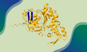
To investigate how the cell’s machinery pulls in the membrane and forms the vesicle, researchers led by Marko Kaksonen and John Briggs employed a method they developed two years ago to faithfully follow the exact same molecules first under a light microscope and then with the higher resolution of an electron microscope. This enabled them to combine two sets of data that so far could only be obtained in isolation: the timing and sequence with which different components of the cell’s machinery arrive at the vesicle-to-be, and the 3D changes to membrane shape that ultimately form that vesicle. They discovered, for instance, that the first proteins to arrive on the inside of the cell’s membrane are not able to start bending it inwards until a network of the cell’s scaffolding protein, actin, forms and starts pulling on the membrane.
The data used to make the video is freely available to the scientific community and will, Kaksonen and Briggs believe, provide valuable information to others trying to develop physical models of how this process works. The EMBL scientists themselves are probing the roles of individual proteins in this process, by perturbing them, and would like to extend the current work in yeast to human cells.
The video accompanying this release is also available on the EMBL YouTube Channel.
Further information:
More work by the Briggs lab on the structure of transport vesicles
Details of the fluorescence and electron microscopy-combining method
Source article
Kukulski, W., Schorb, M., Kaksonen, M. & Briggs, J.A.B. Time-resolved electron tomography reveals how the plasma membrane is reshaped during endocytosis. Cell, 3 August 2012.
Article abstract
Endocytosis, like many dynamic cellular processes, requires precise temporal and spatial orchestration of complex protein machinery to mediate membrane budding. To understand how this machinery works, we directly correlated fluorescence microscopy of key protein pairs with electron tomography. We systematically located 211 endocytic intermediates, assigned each to a specific time window in endocytosis and reconstructed their ultrastructure in 3D. The resulting virtual ultrastructural movie defines the protein-mediated membrane shape changes during endocytosis. It reveals that clathrin is recruited to flat membranes and does not initiate curvature. Membrane invagination begins upon actin network assembly. Subsequently, amphiphysin binding to parallel membrane segments promotes elongation of the invagination into a tubule, which constricts at 1/3 of its depth. Scission occurs on average 9s after initial bending when invaginations are 100 nm deep, releasing non-spherical vesicles with 6’400 nm2 mean surface area. Direct correlation of protein dynamics with ultrastructure provides a quantitative 4D resource.