
The birth of new cells – when two become four
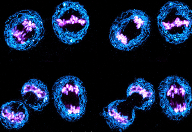
This colourful picture, taken by EMBL postdoc Arina Rybina using a confocal fluorescence microscope, shows human cells in the process of cell division. Eventually, each mother cell brings into existence two identical daughter cells.
To visualise the process by light microscopy, different cell components were labelled using fluorescent dyes. What look like the red teeth of little blue monsters are actually chromosomes. Membranes of a cell organelle called the endoplasmic reticulum are labelled in blue.
The process of cell division has many steps. The chromosomes have to be replicated so that two identical copies of each chromosome are present inside the cell before it divides. The copies align in the centre of the cell. Subsequently, they are dragged to opposite poles of the cell. At the same time, the membranes around each nucleus re-form, to ensure the formation of two fully functional daughter cells.
If you have a stunning picture of your science, your lab or your site, you can submit it here.


