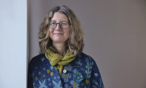
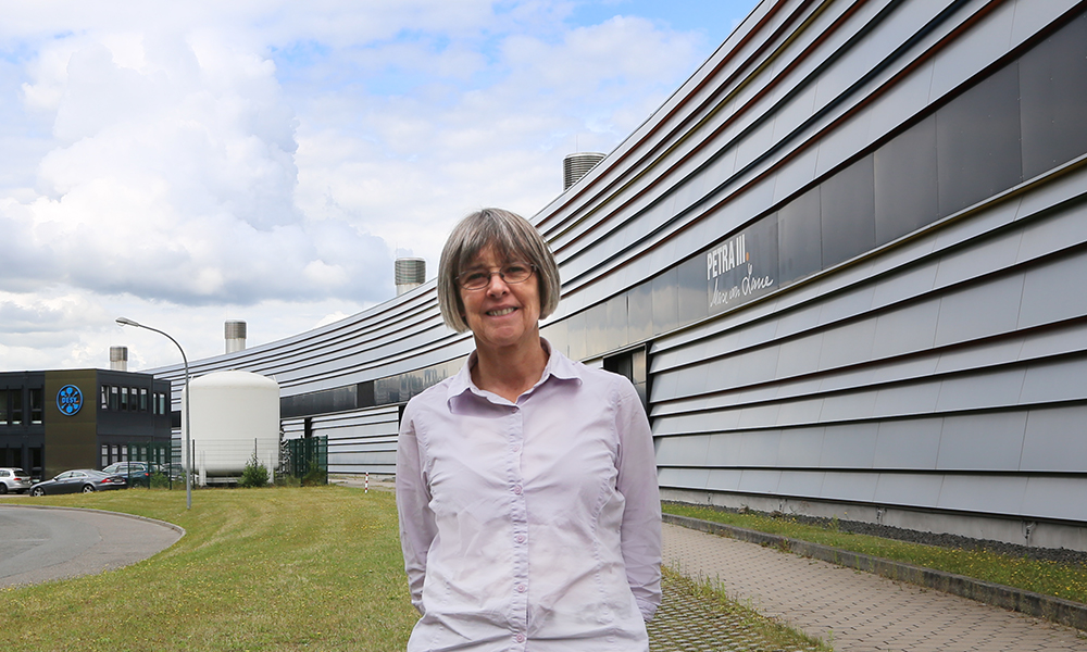
Welcome: Liz Duke
The new team leader at EMBL Hamburg talks about her plans to establish biological X-ray imaging and high-throughput tomography.
Biological X-ray imaging is an emerging technique that uses hard, i.e. high energy, X-rays to image tissues or even entire organisms (in contrast to X-ray crystallography, which is used to probe the structures of proteins and viruses). Biological X-ray imaging will play an important role in EMBL Hamburg’s future portfolio, enabling scientists to answer fundamental questions in biology and to study life on multiple scales.
What is your research background? How did you become interested in science?
My earliest recollection of science was following my father around at CERN when he used to go into the lab at the weekend to catch up on his work. My highlight was when he’d let me feed the punch cards for his computer program into the machine! I was probably about 5 or 6 years old at the time.
My background is physics, but I have always been fascinated by biology. Developing synchrotron X-ray methods that can be applied to solving problems in the life sciences is a perfect way to combine both disciplines. I started my career at the Synchrotron Radiation Source, where I was involved in developing the multiwavelength anomalous dispersion method for protein structure solution. Later, I moved to Diamond Light Source, the new UK synchrotron, where I led the team designing and constructing the first three macromolecular crystallography beamlines. When I started out, most data were collected on film, and beamlines were controlled from the command line. I had the privilege to participate in the development of hardware and software at the Synchrotron Radiation Source and Diamond, which resulted in the macromolecular crystallography beamlines becoming the automated, streamlined facilities that their users have come to expect. Having done that, I wanted a new challenge.
Biology doesn’t happen in isolation. While macromolecular crystallography is incredibly powerful at showing us biological structure at the atomic level, I wanted to explore biological context. This led me to contribute to the development of cryo soft X-ray tomography, a technique that allows you to image the entire volume of a cell without the need for staining or sectioning. I built a beamline, an ‘X-ray microscope’, to do this at Diamond.
Recently, there have been some amazing results using hard X-rays to image thicker, bulk soft tissue samples, and this is my current focus. I want to develop X-ray imaging techniques so that we can image larger samples and use the data to find the tiny region of interest – and thus guide the scientist to where they need to collect the next, higher-resolution dataset.
What motivated you to work at EMBL?
EMBL, and EMBL Hamburg in particular, is a perfect place for me. I have access to the amazing X-ray beamlines, and I am embedded within a community of world-leading biologists who are willing to try out new techniques to advance our understanding of biology. For me, it’s the ideal environment.
What will your team focus on at EMBL?
We will develop new X-ray imaging methods, in particular using hard X-rays to image thicker soft tissue samples. We aim to develop a workflow that will allow us to correlate our X-ray data with data from other imaging modalities, such as fluorescence microscopy and volume electron microscopy.
X-ray imaging at medium resolution can help to pinpoint an area of interest within a sample. This might be a parasite within a zebrafish or a particular cell type within a block of tissue. We are using X-rays to find the needle in the haystack.
X-ray imaging is also perfect for use in neuroscience. Using X-rays to visualise neural pathways means that we can build up neural circuit diagrams relatively quickly and easily.
In the upcoming years, PETRA III, the current synchrotron here in Hamburg, will be upgraded to become PETRA IV. PETRA IV will offer perfect opportunities for biological imaging, so we have some exciting years ahead of us.
A wonderful aspect of being a methods developer is that my work is collaborative. I’m open to work with anyone who is interested in trying out X-ray imaging.
What are some of the challenges of X-ray imaging?
A significant challenge is what to do with all the data we generate. Collecting a dataset takes less than 5 minutes, but processing it takes hours. We need to find ways to speed this up. We have the potential to collect terabytes of data in a day of beamtime. Where are we going to store it all? How do we annotate it so no information is lost? Finally, we want to overlay our data with data from other imaging modalities, and that’s not easy. Fortunately, EMBL has experts in all of this, so I know all these challenges will be tackled.
What do you do when you aren’t X-raying samples?
I have a horse named Indi, and I spend a lot of my time with him. I learned to ride as a ‘mature’ adult, and what I love about it is that it takes my full attention. When I ride, I can’t think about anything other than riding. It’s also great that through horse riding I have been able to meet people from completely different professions and from all backgrounds and walks of life. Indi hasn’t moved to Hamburg yet, but I hope it won’t be too long before we are reunited.