EMBL Imaging Centre welcomes first external user
Correlative microscopy service enables PhD student from Switzerland to study structure and location of proteins cells use to communicate
In September 2021, the EMBL Imaging Centre opened its doors for its first external visitor.
Known for its cutting-edge imaging technologies in the field of light and electron microscopy, EMBL’s Imaging Centre offered Pia Lavriha, a PhD student working at the ETH Zurich and Paul Scherrer Institute in Switzerland, the perfect setting to use both types of microscopes to study how cells communicate.
Lavriha investigates proteins involved in mammalian cell-to-cell communication in the lab of Volodymyr Korkhov. She wants to understand how two cells exchange information with one another in tissues and organs. Her aim is to understand how this process happens on the molecular level.
“My project requires certain techniques and methodologies that are quite complex, and even though I can access the equipment in our lab, I cannot use it in a proper way,” she said. “The Imaging Centre specialists provided the training and insight into how things are supposed to look. I could then develop a feeling for, and expertise in, applying these techniques independently. This is the most important aspect for me.”
Cell-cell communication within tissues is essential to ensure different functions in our bodies happen in sync. Dysregulation of the involved processes can result in a variety of problems including neurodegenerative diseases, developmental changes, or cardiac problems like arrhythmias.
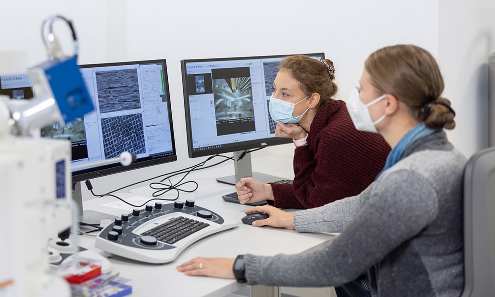
To study the proteins involved in cell-cell communication, Lavriha is combining light microscopy and cryo-electron tomography. The EMBL Imaging Centre offers both technologies in an integrated manner, which enables scientists to study the location as well as the structure of proteins inside cells in detail.
The European access program iNext Discovery, in which EMBL is a partner, supported Lavriha to come to the EMBL Imaging Centre and carry out her project. It is a great example how structural biology facilities can be made accessible to new user communities across national borders.
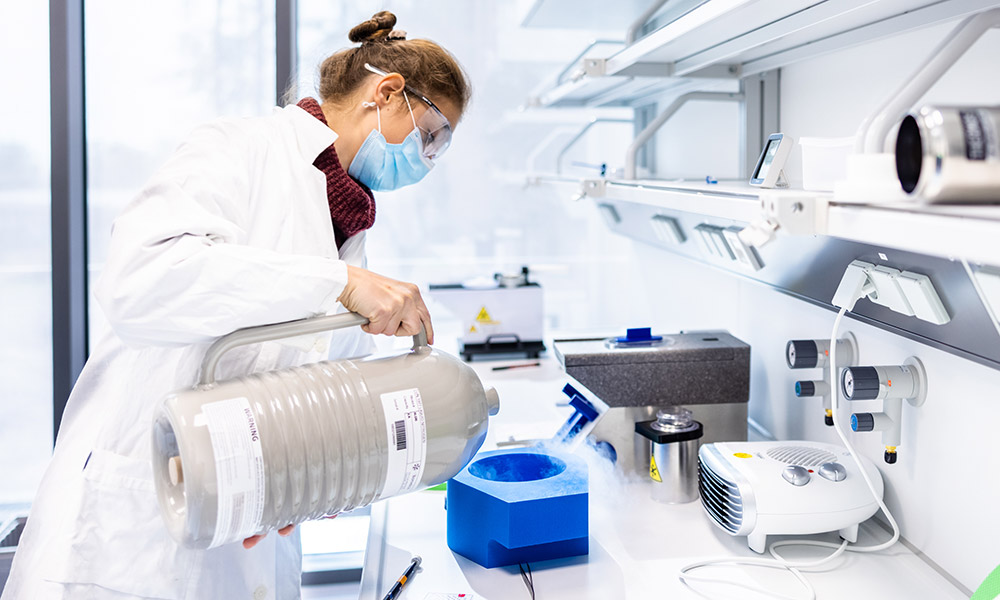
“The scientists at EMBL are among the best in the world in using these techniques,” Lavriha added, explaining how they helped her set up her experimental approach from beginning to end, with expert guidance and training all along the way – from handling samples to interpreting and analysing her results. This training allows her to develop her process independently and later implement it with the machines and equipment available at her home institute.
“The project is designed so that I bring samples and we develop the experimental set-up together. This way I can see how everything works,” she said. “I can then modify the process, so when I visit again I can execute the experiment in the best way possible on my own.”
A call for applications to visit the EMBL Imaging Centre will open soon. More information will be available on the website.