AI and single-cell omics technologies are helping us uncover the rules that determine how cells in glioblastoma – a type of incurable brain cancer – acquire different fates, enabling researchers to experimentally change them
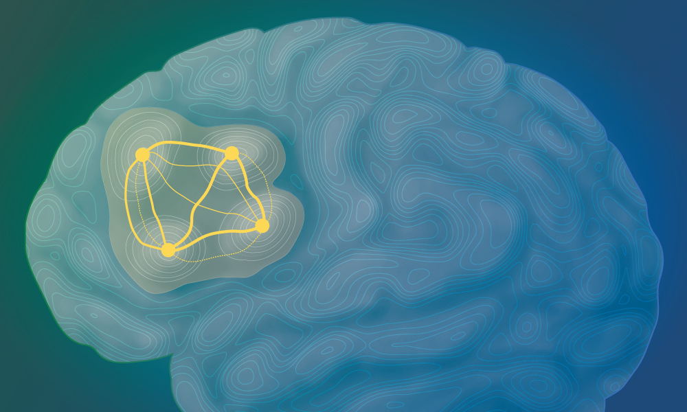
Two new studies demonstrate how glioblastoma tumour cells move between distinct cellular states and how this process is regulated. Credit: Maria Oliveira/EMBL
Summary
Glioblastoma kills over 100,000 people each year worldwide and is characterised by tumour cells that take on a range of cellular states.
In two companion studies, researchers have shown that these cells follow predictable and repeatable paths between such states, and that this process is governed by distinct gene regulatory networks.
The researchers were also able to systematically reprogram these states, pushing cells in a less malignant direction, increasing survival in mouse models.
These new findings help us better understand how tumour cells evade treatment, and could identify molecular targets for more effective therapies in the future.
Glioblastoma is the deadliest and most common type of malignant brain tumour in adults. One of the reasons it’s almost always incurable is that individual tumour cells are very different from one another and shift dynamically between various ‘states’. This plasticity helps them evade anti-cancer treatments.
Now, two new studies from researchers at EMBL, the German Cancer Research Center (DKFZ), and the Wellcome Sanger Institute, UK, show that this state-shifting follows predictable patterns, providing hope for future therapies targeting these cancers. The results were recently published in the journal Nature.
Building a molecular atlas of glioblastoma cells
Researchers and clinicians have long been stymied by the high heterogeneity of glioblastoma tumours. Cells within these tumours exist in a variety of states, each with distinct molecular features. Some resemble nerve cells, while others look like stem cells or become trapped in an aggressive, injury-like state. The cells can also transition from one state to another, a phenomenon known as plasticity. Since therapies may target some cell states but not others, such plasticity helps the cancer cells survive treatment.
However, the prevalent view in the field has been that such state-shifting is essentially random and very difficult to predict. This is the view the researchers in the current study challenged by creating and analysing one of the most comprehensive molecular datasets derived from glioblastoma patients to date.
“We asked – can we go back to the drawing board?” said Omer Bayraktar, Group Leader at the Wellcome Sanger Institute and a new professor at the UCL Cancer Institute. Bayraktar’s team uses single-cell spatial technologies to study the cellular diversity of brain disorders. “Can we assume that we know nothing about this disease, and reconstruct how a cancer cell behaves in glioblastoma through quantitative approaches?”
Funded by a Wellcome Leap grant, Bayraktar’s team examined DNA, epigenetic marks (a type of molecular ‘annotation’ of genes that regulates their expression), and RNA in tumours from 12 patients, generating the largest glioblastoma molecular atlas to date. To make sense of this highly complex dataset, they teamed up with Oliver Stegle’s team at EMBL Heidelberg and DKFZ, who are experts in applying advanced computational approaches to fundamental life science problems.
“We wanted to figure out whether there is a specific rule-based system that determines how cells transition between these states,” said Stegle. “And we asked not only whether there were rules, but also what would drive such rules.”
Together, the researchers found that, contrary to what was known before, glioblastoma cells followed certain stereotypical, predictable paths between cell states. Different cell states also occupy different spatial niches inside a tumour.
“By integrating the single-cell data with spatial transcriptomic data, we found that there is a meticulous and very specific spatial architecture of cancer cell states,” explained Bayraktar. “And this was present in every single glioblastoma tumour in every single patient we studied.”
The team further confirmed this data using organoids – tumour cells taken from patients and grown in the laboratory on a three-dimensional scaffold, closely mirroring conditions inside the brain. They found that similar cell state trajectories emerged in patient-derived organoids as what they had deduced earlier from the glioblastoma atlas data.
Understanding the rules and regulators that govern state transitions
Now knowing that glioblastoma cells follow certain stereotypical trajectories between states, the researchers then set out to investigate how this process is regulated. For this, they turned their attention to gene regulatory networks. These are the rules and molecular regulators that determine which genes are expressed in which cells and when.
Led by Moritz Mall’s team at DKFZ, the researchers examined gene regulation signatures in over 1 million patient-derived glioblastoma cells and used this to figure out how tumour cells transition between states. They identified a structured regulatory program that governs cellular plasticity in these cells, one that was both predictable and hierarchical.
The team also identified a set of ‘safeguards’ – regulators which arrest cells in low-plasticity states, preventing them from transitioning to other states. This was of particular interest as these regulators could potentially be targeted genetically or pharmacologically as part of cancer therapies in the future.
“One of our premises has always been that when we understand these rules, we can not only observe what the cells are doing, but we can also change what they are doing,” said Mall. “We could perhaps reprogram them, and maybe push them into a state that is less malignant, less dangerous.”
The researchers tested this hypothesis in patient-derived brain tumour cells, where they found that overexpressing MYT1L, one such ‘safeguard’ regulator identified by this study, could reduce cellular plasticity and tumour malignancy. If transplanted into mice, these ‘modified’ tumours resulted in better survival than control tumours.
“The results with MYT1L were interesting, but the findings go much beyond that,” explained Mall. “Our approach predicts the regulators for all of the major tumour cell states, and we tested those predictions experimentally. We were able to steer tumour cells into specific states by manipulating just a handful of regulators.” In other words, the predictions of the new model could be used to engineer the identity of a tumour cell.
AI turns tumour diversity into a source of insight
The diversity that makes glioblastoma so hard to treat also turned out to be the key to understanding it. Each tumour, and each cell within it, carries its own combination of genetic and epigenetic changes. Across a million cells, this adds up to a vast number of natural variations in how genes are regulated, far more than researchers could ever create in the lab.
To learn from this variation, Stegle’s team developed scDORI, a deep learning method that infers gene regulatory networks at single-cell resolution. By comparing how regulators and their target genes change together across more than a million cells, scDORI pinpoints which regulators steer cells towards one state or another. These predictions were the starting point for the reprogramming experiments carried out by Mall’s team.
“A tumour generates an almost endless number of variations on its own regulatory programme,” said Stegle. “Instead of seeing this heterogeneity only as an obstacle, we can use AI to learn from it and extract the rules that determine how cells choose their fate.”
A second machine-learning method mapped where each cell state sits within the tumour, revealing the spatial architecture described above.
The researchers are now applying this approach to other cancers and brain disorders. While clinical applications may still be some way off, this is already helping researchers understand tumour cells at the deep molecular level and spot common patterns. “With the help of this atlas and the new AI tool, we identified new regulatory sequences that enable us to potentially drive specific cell states in a tumour-specific setting,” said Mall. “Now that we have this framework, we can apply it to many other questions and cancers.”