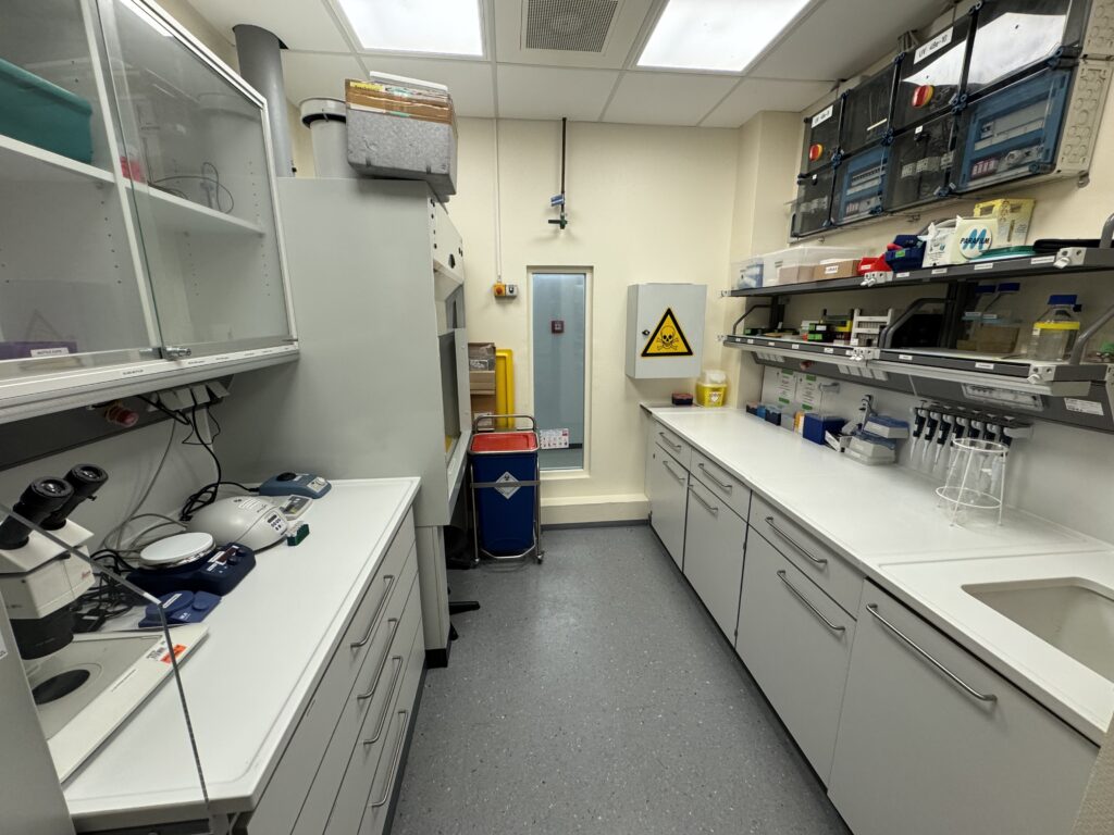
Sample Preparation Laboratory
Here you can find the present equipment and chemicals that are available on site in the sample preparation laboratory.
High-Throughput Tomography (HiTT) on P14
Below, you can find all the necessary information for successfully preparing your samples for measurement at our beamline.
Following positive evaluation of your proposal, you will receive an invitation letter for a scheduled beamtime session which has been allocated to your project.
Please make sure that you accept the scheduled beamtime before the deadline stated in the invitation.
To enable a smooth visit to our facility, we encourage you to read the following instructions and information carefully. In case of any doubt or if you have questions, please do not hesitate to contact the Duke Team or the EMBL User Office.
Below, you will find all important information and links about:
In order to increase the likelihood of collecting high-quality data, we recommend that your samples be prepared following the detailed instructions provided here. It is important to stay within the sample sizes given and to follow the instructions explained below.
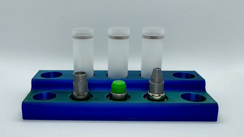
Our established and standardised protocols enable the acquisition of X-ray imaging data from paraffin-embedded samples, resin-embedded samples, and samples in liquid. The samples are mounted on SPINE-style pins that sit in a puck, which enables fully automatic sample handling (see SPINE sample mounting system).
Please note that the final stages of sample preparation and mounting can be conducted in a laboratory facility adjacent to the beamline, which is equipped with all necessary liquids and materials. However, more extensive preparatory processes, such as dehydration, resin embedding, and paraffin embedding, are not available within our laboratory premises.
The sample preparation process for your specific samples may differ from the general guidelines outlined below. The Duke Team is dedicated to assisting you with your unique requirements and the detailed preparation of individual samples. We look forward to providing tailored support to meet your needs.
Here you can find the present equipment and chemicals that are available on site in the sample preparation laboratory.
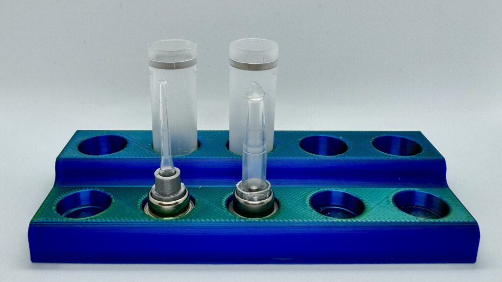
Liquid samples should ideally be scanned in ethanol to ensure optimal results. We have developed specialised adapters to facilitate the connection of sealed pipette tips to the SPINE base. For larger samples, it may be necessary to stitch together multiple scans (see sample sizes and acquisition times). In such instances, it is crucial to maintain a concentration of at least 70% ethanol to enable smooth acquisition and minimise sample movement due to radiation-related bubble formation. If a single scan is sufficient, samples can also be scanned when mounted in 50-70% ethanol or paraformaldehyde (PFA), although this method carries a higher risk of movement during scanning, potentially leading to unusable data.
It is important to note that any variation in the liquid sample mounting process can lead to complications during acquisition. Therefore, we strongly advise carrying out the mounting of liquid samples on-site in the designated sample preparation laboratory on the day preceding the scheduled beamtime in order to allow the sample to settle and any bubbles created during the mounting process to disperse.
For a detailed explanation of the sample preparation process for liquid samples, please refer to the accompanying sample preparation video.
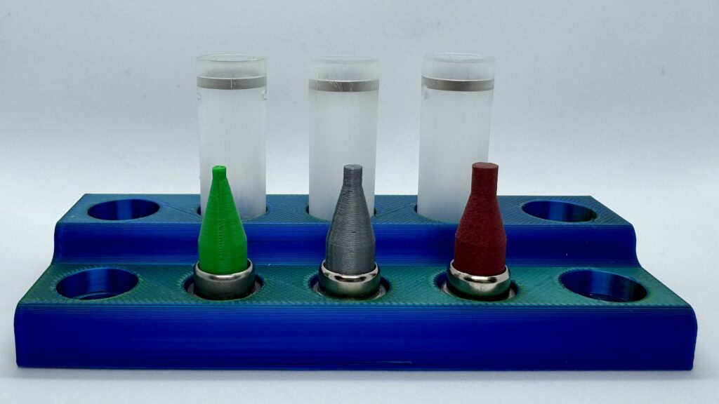
Resin embedded samples can be effectively analysed at our beamline utilising a specialised sample mount that has been adapted for standard EM-resin blocks. The design of the variable adapter enables customisation to meet the specific requirements of the user. This adapter integrates seamlessly with the SPINE base through a plug-and-play mechanism, enabling efficient attachment. The resin block is securely affixed to the adapter using an adhesive, and it can be detached without causing any damage, thus permitting its use in subsequent applications.For a detailed explanation of the sample preparation process for liquid samples, please refer to the accompanying sample preparation video.
Formalin fixed paraffin embedded (FFPE) samples can be scanned at the beamline, The best data can be obtained if the samples are of cylindrical shape (e.g., produced by a punch biopsy). The punches can be glued on specially designed paraffin mounts that serve as an adapter to the SPINE bases. The available adapters accommodate cylindrical FFPE samples ranging from 1 mm to 3.5 mm in diameter, with a maximum length of 1.2 cm. Adapter pins can be provided and sent to users upon request, or they can be utilised and affixed in the sample preparation laboratory on-site, prior to the commencement of beamtime.
For a detailed explanation of the sample preparation process for liquid samples, please refer to the accompanying sample preparation video.