Get in touch
Contact the Duke Team for further information.
High-Throughput Tomography (HiTT) on P14
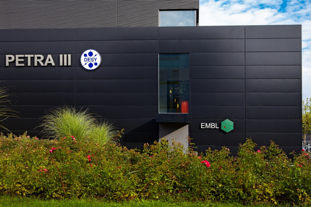
The X-Ray Imaging Team, led by Liz Duke, provide a synchrotron-based X-ray micro-tomography facility for biological samples. We are located at the EMBL P14 beamline of the PETRA III synchrotron light source, on the DESY campus in Hamburg (Germany).
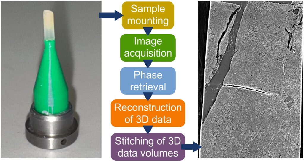
Our setup, which we call High-Throughput Tomography (HiTT), uses synchrotron X-rays to perform high-resolution scans of samples of sizes in the millimetre range. The high intensity of the X-rays makes it possible to image at a high speed: for samples below about 2 mm in size, data acquisition and reconstruction of the 3D data takes less than five minutes, which, because we also use a robot, enables us to scan a large number of samples in a moderate amount of time.
Experiments involving X-ray soft imaging of biological tissue can be performed at the EMBL beamline P14, at the PETRA III storage ring at DESY (Hamburg, Germany), coordinated by the X-ray Imaging Team led by Liz Duke. Users can apply specifically for X-ray imaging (XIMG) beamtime via the EMBL Hamburg beamtime application process.
Contact the Duke Team for further information.
Find useful information on how to submit your beamtime proposal, prepare your visit, or receive access funding.
Thank you very much for joining our first online user meeting. If you could not attend live or want to take another look at the presentations, you will find the slides from our presenters and recordings of the presentations below.
If you have further questions for the presenter, please use the contact button on the right to get in touch with the Duke Team.
The presentation gives an overview of the application for X-ray imaging beamtime at the EMBL Beamline P14. Liz will guide you through the different requirements for a successful application for beamtime. If you have further questions, please get in touch with the Duke Team. Below you can download the presentation slides.
The presentation overviews the High-throughput Imaging technique and setup used at P14. Jonas will guide you through the principles of X-ray imaging, X-ray tomography, and reconstruction. Further, he will talk about the updates and improvements for the users. Below you can download the presentation slides.
The presentation will give you a detailed overview of the established sample types that can be scanned at the EMBL P14 beamline. Luaks will show you some tips and tricks to improve sample preparation and mounting. Further, he will talk about the on-site sample preparation laboratory, which users can use.
Below you can download the presentation slides.
This presentation introduces Webknossos by Scalable Minds, an open-source tool for annotating and exploring large 3D image datasets in life sciences. Norman will guide you through the different tools, including segmentation, web-based browsing, AI implementation, etc.
This presentation gives an overview of how the tool Webknossos was recently implemented into the workflow of HiTT imaging at EMBL Hamburg to create a remote data visualization and annotation platform for our users.
This presentation will give you an overview of the postdoctoral project of our former postdoctoral Fellow, Matthew Lawson, who is now a Senior Technical Specialist (NXCT) at the University of Manchester. He will explain his work as part of the Medical Research Council (MRC) to acquire multimodal imaging data of human kidney samples on a large scale. Below you can download the presentation slides.
This presentation will give you an overview of Fabio’s project, which applies an alternative imaging method called Speckle-based imaging at our EMBL Beamline P14 in Hamburg. This technique makes it possible to receive quantitative grey values from the investigated tissue. Below you can download the presentation slides.