Edit

Protein Expression and Purification Core Facility

PEPCF expresses proteins in bacteria, insect and mammalian cells and uses a variety of chromatographic and biophysical techniques for protein purification and characterization.

Biophysical support

The biophysics platform provides support for the design, execution and data analysis of biophysical and biochemical experiments. We aim to assist users in characterizing their proteins and also ensuring that proteins are of high quality and suitable for downstream experimentation.

Within the biophysics platform, we have many techniques for characterizing your proteins and protein complexes. We have capacities for analyzing interactions (isothermal titration calorimetry, microscale thermophoresis, fluorescence polarization and surface plasmon resonance), determining molecular weight of samples such as oligomerized proteins, protein complexes, and modified proteins (SEC-MALS, mass photometry and DLS), finding optimized buffer conditions and protein thermal stability (nanoDSF and thermofluor), determining secondary structure (circular dischroism), and for overall protein quality control to help ensure downstream experiments reflect high quality experimentation using biologically relevant proteins.

These experiments can be performed by the PEPCF scientific officer for biophysical support or the users can be trained to execute and analyze their own experiments independently. Please contact Karine Lapouge to discuss experimental specifics and sample requirements.

We have three different types of spectrometers available for use in our facility: a circular dichroism spectrometer (Jasco J-815), two fluorescence spectrometers equipped for both standard fluorescence and fluorescence polarization (Jasco FP-8500 (cuvette) and plate reader BioTek Synergy 4), and two UV-Vis spectrometers (Agilent Cary 60 (cuvette) and plate reader BioTek Synergy 4).


Isothermal Titration Calorimetry

Isothermal Titration Calorimetry (ITC) is a label-free thermodynamic technique that measures the heat produced or consumed during a binding interaction. A ligand in the syringe is titrated into the macromolecule in the cell. Measurement of the temperature change, relative to the reference cell, allows for accurate determination of binding constants, reaction stoichiometry and a complete thermodynamic profile for the interaction.

Sample requirements: usually starts with concentrations of 20 µM (350 µl) for the sample in the cell and 200 µM (75 µl) for the titrant sample in the syringe. These concentrations are used as starting conditions producing information for optimization when approximate binding constants are unknown. When the Kd is estimated for a 1:1 binding, the following recommendations for sample concentrations can be applied.

MicroCal PEAQ ITC from Malvern Panalytical

scientific machine

MicroScale Thermophoresis

Microscale Thermophoresis (MST) measures biomolecular interactions between a fluorescently labelled sample and a binding partner (protein, RNA, DNA, small molecule etc.) by recording changes of fluorescence intensity when a thermal gradient is applied to the sample.

Sample requirements: approximately 20 nM of the labelled analyte and varied concentrations of the ligand spanning a full range around the Kd (typically 0.01-20 x Kd). Options for labelling include for example NHS coupling, thiol coupling and His-tag labeling. Each capillary requires 12 µl, and a single experiment usually requires >26 capillaries (2 for initial fluorescence check, 8 for initial binding check, and 16 for a single titration series). Optimization is often required to find the best conditions.

Monolith NT.115 from Nanotemper


SEC-MALS

Multi-angle light scattering (MALS) coupled to size exclusion chromatography (SEC) monitors the absolute molecular weight of proteins or macromolecular complexes independent of the SEC elution profile.

The Wyatt miniDAWN and Optilab instruments measure static light scattering and differential refractive index (dRI), respectively, for determining particle molar mass and concentration. We have an Agilent HPLC system set up together with our Wyatt detectors for loading the samples. The SEC columns we have available for sample separation are a Superdex 200 Increase 10/300 GL, a Superdex 200 Increase 5/150 GL, a Superdex 75 Increase 10/300 GL and a Superose 6 Increase 10/300 GL. This instrumentation can be cooled to 10ºC. Sample runs and analysis are performed using the Wyatt Astra software version 7.

SEC-MALS is routinely used for determining the absolute molar mass of protein samples and is an important component of protein quality control.

Sample requirements: approximately 50-500 µg of sample (10-100 µl can be injected; 10 µl should be added for the autosampler dead volume).

HPLC from Agilent – Mini Dawn detector, Optilab detector from Wyatt


Thermal Shift Assays

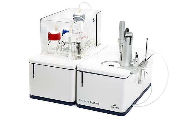
Protein thermal stability can be analysed using thermal shift assays. The thermal stability of a protein is influenced both by the nature of the protein and its chemical environment (i.e. pH, salt composition and concentration, additives and the presence of various ligands) and changes can be determined by observing shifts in the protein melting temperature (Tm). Thermal shift assays are used for determining the optimal conditions for stabilizing proteins and screening of potential interactions between a protein and ligands.

We use two different techniques for performing thermal shift assays, which are Thermofluor and nano-Differential Scanning Fluorimetry (nano-DSF).

We perform our Thermofluor assays in a real-time PCR instrument. In thermofluor assays, a dye (e.g. SYPRO orange) that fluoresces upon binding to hydrophobic patches is added to the samples. As the sample is heated up over time, the protein will start to unfold and expose more hydrophobic patches, which causes an increase in fluorescence. Monitoring the change in fluorescence over time is used to determine the melting temperature Tm.

For measuring thermal stability with nano-DSF, no fluorescent dye is required. The samples are loaded in capillaries and changes in intrinsic fluorescence (Trp, Tyr) are monitored over time while the samples are heated up. Our instrument is also equipped with back-scattering optics, which allows detection of sample aggregation as well. Nano-DSF is also routinely used as a quick protein quality control step.

For determining optimal buffer conditions for protein stability, we have both the RUBIC Buffer screen and RUBIC Additive screen from Molecular Dimensions available.

Thermofluor sample requirements: protein concentration ~ 20 µM; 2 µl sample per well in a 96-well plate.


Nano-DSF sample requirements: protein concentration ~ 5 µg/ml – 150 mg/ml, dependent on the Trp – Tyr content of the protein; 10 µl of sample per capillary.

CFX Connect Real Time PCR system from Bio-Rad (thermofluor)

Prometheus NT.48 with backscattering optics from Nanotemper (nano-DSF)


Surface Plasmon Resonance

Surface plasmon resonance (SPR) monitors changes in mass concentration at the surface of the sensor chip. A ligand is immobilized on the surface of a sensor chip and interacts with an analyte injected over the sensor surface. SPR can be used to measure real-time biomolecular binding affinities and kinetics.

The Biacore T200 instrument is located in the EMBL Chemical Biology Core Facility (CBCF) and more information about this service can be found on the CBCF website.

Biacore T200 from Cytiva


Dynamic Light Scattering

Dynamic light scattering (DLS) measures the time-dependent fluctuation of scattered light intensity caused by the Brownian motion of particles in solution. It can be used to determine particle size and to assess the homogeneity and oligomeric state of macromolecules. DLS is commonly used for protein quality control as well.

Sample requirements: approximately 80 µl of a 0.5-2 mg/ml sample.

Zetasizer µV from Malvern


Mass Photometry

Mass photometry measures the molecular mass of label-free single molecules in solution, by quantifying light scattering from individual biomolecules in solution. 

The mass photometry technology is based on the principles of interference reflection microscopy and interferometric scattering microscopy. The interference between the light scattered by a molecule upon landing on a glass surface and the light reflected at the glass-water interface is measured. The resulting mass photometry contrast correlates linearly with molecular mass.

It can be used for protein quality control, determination of sample purity and homogeneity, protein oligomeric state, and characterisation of biomolecular complexes assembly.

Sample requirements: approximately 20 µl of sample at a concentration of 400 nM. Buffers should be filtered (0.22 µm) prior to measurement and high glycerol concentration (>10%) should be avoided. Mass range: 30kDa – 5MDa.

Refeyn TwoMP from Refeyn


As the topics investigated by researchers in the life sciences become more and more complex, an interdisciplinary and collaborative approach is often required to successfully address these complicated biological problems. Recombinant proteins are popular tools that are frequently used to investigate many aspects of these biological questions by researchers from different scientific backgrounds. At the EMBL Protein Expression and Purification Core Facility we work together with scientists from various fields, ranging from structural biology to cell biology to medicine and pharmaceutical research.

Over the years we have learned that the conditions required for recombinant proteins vary strongly between all of these fields. For example, proteins used for structural biology by a crystallographer or an electron microscopist have different concentration and buffer requirements, and these vary greatly from protein conditions used for in vivo experiments by medical researchers.

Edit