Focused Ion Beam Scanning Electron Microscope (FIB-SEM)
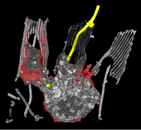
The Focused Ion Beam – Scanning Electron Microscope (FIB-SEM) is an instrument that allows the imaging of a sample in 3D at nanometer resolution in a semi-automated way. It combines iterative imaging with a SEM and physical slicing of the sample with a Gallium ion beam. The iteration of imaging and milling generates a stack of images that are then digitally combined to reconstruct a 3D volume.
In the EMCF we are operating 2 FIB-SEMs, a Zeiss CrossBeam 540 and a 550.
With these microscopes we can acquire datasets at high (isotropic) resolution, up to 5 nm voxel size. Nevertheless, the target volume is limited to a few (tens) of cubic micrometers, which allows us to acquire from subcellular volumes to single cells or small tissues. Examples of samples recently acquired in the EMCF are sponge cells, drosophila tissues, organoids, cultured mammalian and yeast cells, mouse tissues.