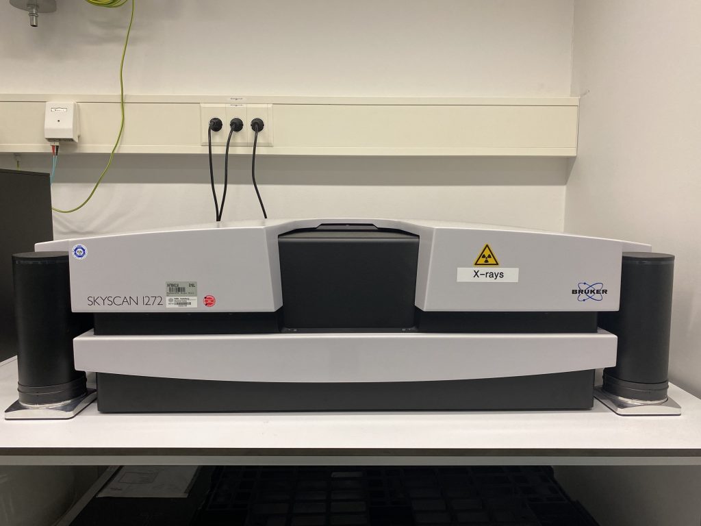
Use of the Skyscan 1272 microCT has been heavily embedded into our 3D CLEM workflows in the EMCF. Samples are fixed, by means of chemical fixation or high pressure freezing/freeze substitution, incubated with osmium tetroxide and heavy metals (introducing contrast to the sample) and embedded into a resin where it will be either imaged in the TEM or SEM. Once the resin block is scanned, we are left with a 3D map of the sample. We can measure the distance from the block surface to the ROI and trim away the material in-between bringing the ROI directly to the surface for imaging.
Reference:
Karreman MA, Ruthensteiner B, Mercier L, Schieber NL, Solecki G, Winkler F, Goetz JG, Schwab Y, Find your way with X-Ray: Using microCT to correlate in vivo imaging with 3D electron microscopy. Methods Cell Biol. 2017:140:277-301.