The Scanning Electron Microscope (SEM) allows one to explore the surface of a specimen, to better understand its topology. Specimens ranging in many sizes can be processed and scanned using this method.
Specimen processing begins with a mild chemical fixation followed by a post-fixation with heavy metals to obtain contrast. To withstand the high vacuum in the SEM, water molecules within the sample are removed (with the addition of ethanol or acetone) and the sample is completely dried while preserving the surface topography. This is achieved by adding an hydrophobic HMDS layer on the surface of the specimen or by critical-point-drying (CPD).
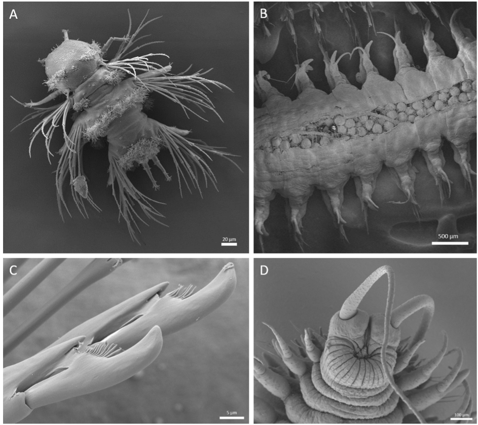
We are imaging using a Zeiss Crossbeam 540. The surface of the specimen is scanned with the electron beam down to nanometer resolution. An image is formed by detecting the secondary electrons emitted from the sample surface or from the backscattered electrons.The EMCF and its users have studied the topology of many diverse samples with SEM such as mouse embryonic stem cells (Bergert et al. 2020), mouse liver, Platynereis dumerilii and marine plankton.
References:
Bergert M, Lembo S, Sharma S, Russo L, Milovanović D, Gretarsson KH, Börmel M, Neveu PA, Hackett JA, Petsalaki E, Diz-Muñoz A. Cell Surface Mechanics Gate Embryonic Stem Cell Differentiation. Cell Stem Cell. 2021 Feb 4;28(2):209-216.