Edit

Advanced Light Microscopy Facility

This facility offers a collection of state-of-the-art light microscopy equipment and image processing tools.

Services

We encourage prospective users and their supervisors to join an initial project discussion to clarify microscopy needs to ensure optimal imaging guidance.

The ALMF offers the following microscopy and high-throughput services:

Image courtesy: Annika Vogler,
Furlong group

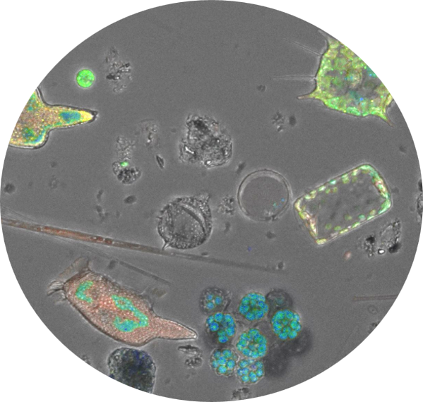
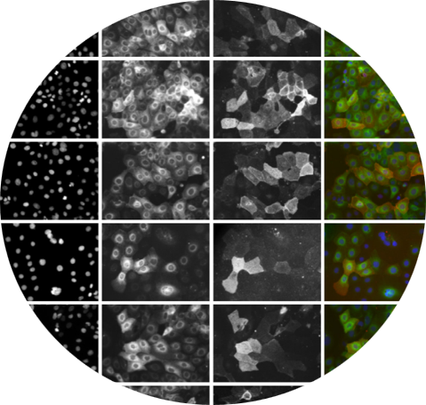
Widefield microscopy

Wide-field microscopy uses visible light to capture images of whole samples quickly, without rejecting out-of-focus light. It is great for observing live cells and large areas. It’s a key tool for general observation and initial assessments.
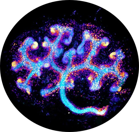
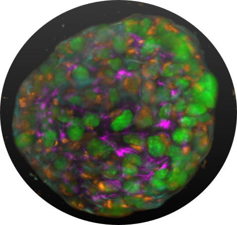
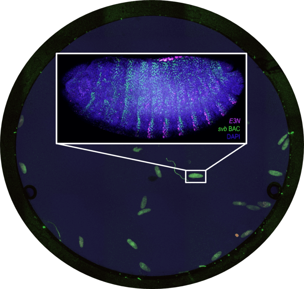
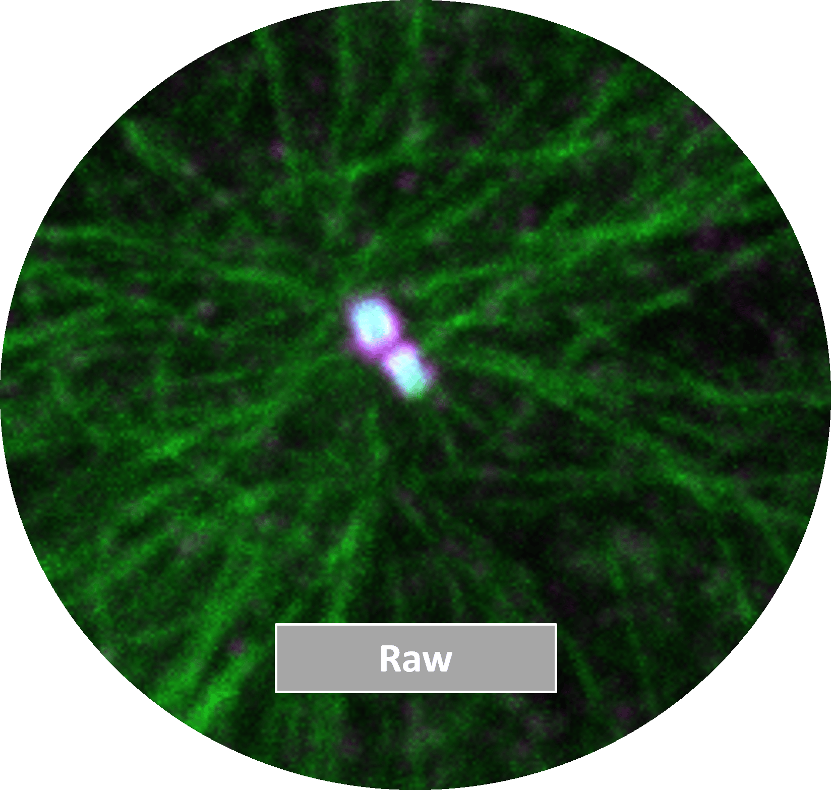
Explore systems

Image courtesy: Nobuko Tsuchida-Straeten, Simons & Aulehla lab

Confocal microscopy

Confocal microscopes capture optical slices by rejecting out-of-focus fluorescence, enabling the acquisition of z-stacks for precise 3D imaging. It provides high contrast and clarity, making it ideal for studying complex biological processes.

Explore systems

Additional modalities available:

Spinning disc confocal

AIRY fast confocal

Sample courtesy: Enrico Salvatore Atorino, Banterle & Dodonova labs, imaged by ALMF

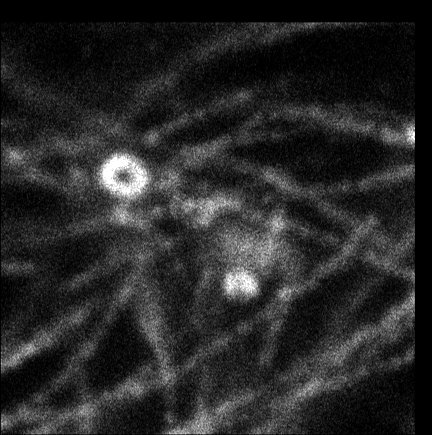
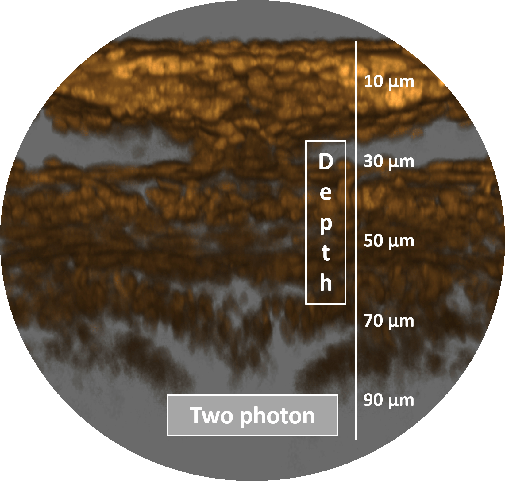
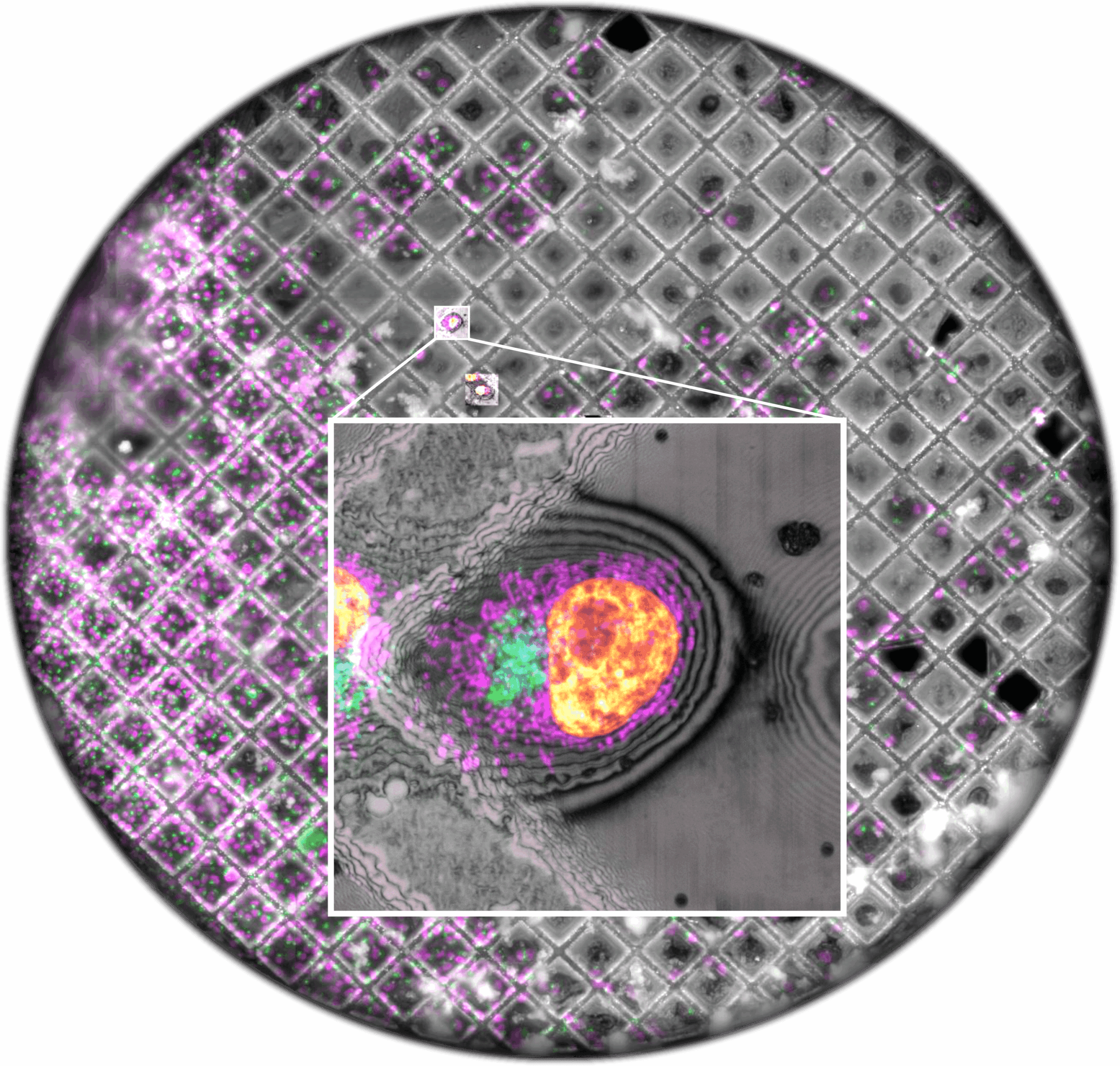
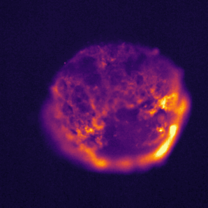
Super-Resolution microscopy

Super-resolution microscopy uses techniques like Single Molecule Localization Microscopy (SMLM) and Stimulated Emission Depletion (STED) to go beyond the diffraction limit of light, providing high-resolution imaging at the nanometer scale. With achievable resolution down to ~20nm, this technique lets us study biological processes at the molecular level, bridging light and electron microscopy.

Explore systems

Sample courtesy: DB unit, imaged by ALMF

Multiphoton microscopy

Multiphoton microscopy uses long-wavelength light to excite fluorescence only in the focal volume. This reduces scattering and photobleaching, making it ideal for imaging thick, living samples with less photodamage. The technique is especially effective for deep tissue imaging.

Explore systems

Sample courtesy: Schwab group, imaged by ALMF

F-techniques

F-techniques like FRET, FRAP, FCS, and FLIM are used to study molecular interactions, particle movement, diffusion, and fluorescence dynamics in live cells, providing detailed insights into cellular processes and molecular environments.

Explore systems

Sample courtesy: Martin Jechlinger, imaged by ALMF

Lightsheet microscopy

Light-sheet microscopy uses thin light sheets for fast, high-contrast 3D imaging with minimal photodamage, making it ideal for imaging large, live specimens.

Explore systems

Image courtesy: Aliaksandr Halavatyi, ALMF

Advanced workflows

Feedback microscopy

Optogenetic microscopy

Laser micro-dissection microscopy

RNAi technology

Image courtesy: Martin Fritsch, ALMF

Cross disciplinary workflows

Correlative light and electron microscopy (CLEM) with EMCF /Imaging Centre

Drug screening with Chemical Biology Core Facility (CBCF)

Image courtesy: Pinto et al., 2022, J. Molbiol

High Throughput microscopy

Automated imaging approach that allows rapid screening and analysis of large numbers of samples or cells. It combines robotics, advanced imaging, and data processing to efficiently handle large-scale experiments, such as drug screening, genetic screens, or cell behavior studies.

Explore systems

Sample courtesy: Mereke Suleimenov, Banterle lab, imaged by ALMF

Data processing, handling & management

For more advanced analysis, contact BiAS

We offer support for basic image handling (Fiji), Deconvolution (Huygens), 3D visualisation/segmentation (Imaris) and access to virtual image processing machines

Edit