3D-SMLM
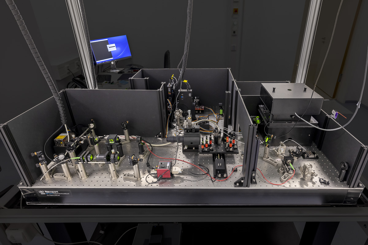
Three-dimensional single-molecule localisation microscopy (3D-SMLM) is an advanced light microscopy methodology based on localisation of sparse single-molecule emitters for computational reconstruction of a super-resolved image. The 3D-SMLM at the EMBL IC has been developed in collaboration with the lab of Jonas Ries at EMBL. At the core of the microscope is an extremely stable inverted microscope. (f)PALM/(d)STORM workflows can be accommodated and additional functionalities will be added dependent on user need (e.g. support for live-cell imaging/PAINT/SOFI and others).
Features
- Ratiometric multi-colour imaging (e.g. simultaneous multi-colour imaging of spectrally overlapping fluorophores)
- Homogenised Epi-/HILO/TIRF illumination with variable illuminated field ca. ⌀20 – 70 microns, Super-resolution over large field of views (up to ⌀70 microns)
- Option for 3D imaging via astigmatism
- < 10 nm localisation precision (in x,y, dependent on fluorophore, imaging mode, < 20 nm in z)
- Automated high-throughput imaging (e.g. multiple field of views)
Specifications
- 4 lasers generating ca. 100 mW at source (Toptica iChrome MLE, 405, 488, 561, 640 nm), additional booster laser at 640 nm (Toptica iBeam Smart 200 mW).
- Back-thinned sCMOS camera (Hamamatsu Orca Fusion BT >95% QE, <0.7 e- read noise)
- Focus locking via reflection of NIR laser (Toptica iBeam Smart, 785 nm)
- Motorized XY nanopositioning stage (SmarAct), Piezo actuator (PI PIFOC, P-726) for objective.
- Olympus 100x/1.45 (oil), 100x/1.35 (silicone oil) objective lenses
- The microscope can be configured for the needs of the experiment via a range of switchable optical elements. 3D imaging is achieved using the astigmatic method and analysis/reconstruction is achieved using the SMAP toolset
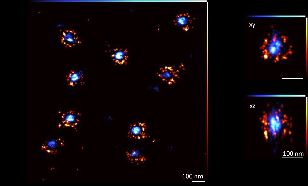
that the two stacked rings can be resolved. Scale bars, 100 nm. Credit: Merle Hantsche-Grininger/EMBL.