New services in 2026 – Serialised lift-out for cellular tomography
This year, we are expanding our portfolio with three powerful new imaging services, giving the life-science community access to cutting-edge technologies for studying biological structure, dynamics, and mechanics across scales. Serialised lift-out for cryo-electron tomography, Oblique Plane Microscopy (OPM), and Brillouin Microscopy — are designed to address some of the most challenging questions in modern biology by enabling imaging in near-native conditions, at high resolution, and with minimal perturbation to living systems.
Serialised lift-out for cellular cryo-electron tomography
From native tissue to molecular architecture
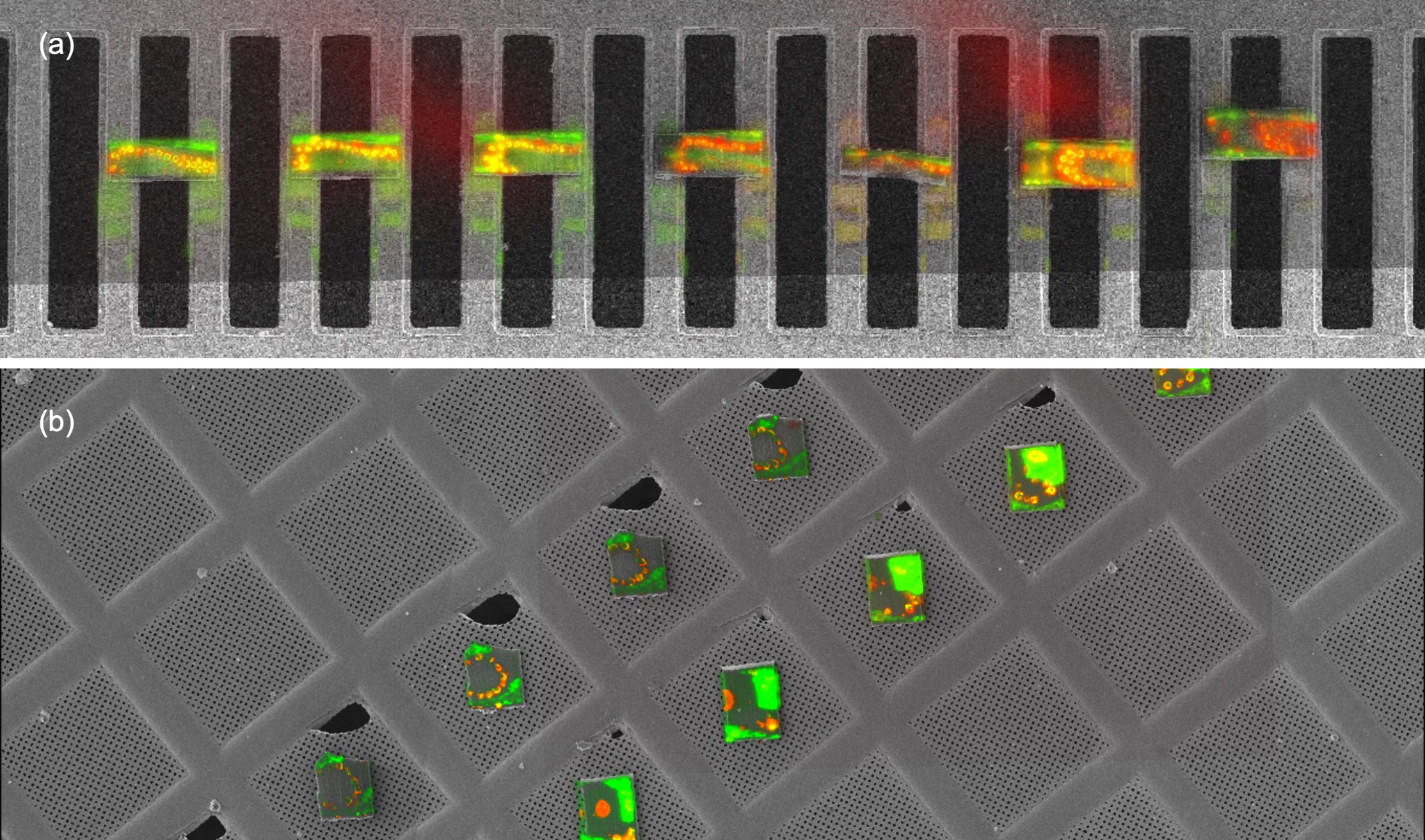
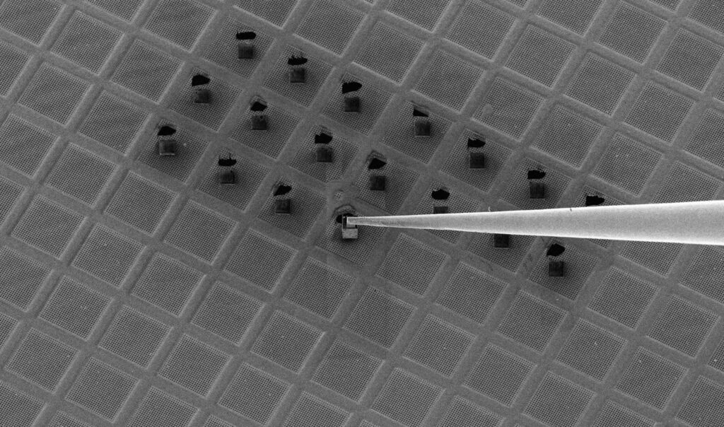
The first new service now available to the wider research community is ‘serialised lift-out’’. This transformative cryo-focused ion beam (cryo-FIB) technique enables high-resolution cryo-electron tomography (cryo-ET) of thick, complex biological samples, including tissues and whole organisms that can be vitrified by high-pressure freezing (HPF). Building on recent methodological advances established by the Plitzko and Erdmann labs [1,2], this approach allows multiple thin lamellae to be extracted sequentially from a single vitrified specimen, preserving native cellular context while dramatically increasing throughput.
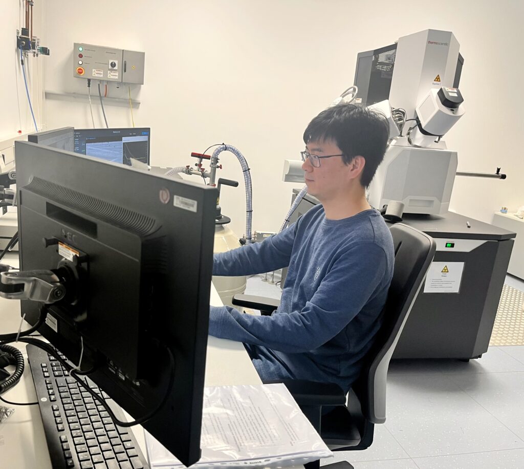
Already in 2024, the cryo-EM team began evaluating serial lift-out in response to its strong potential for life-science projects and broad range of applications. Early implementation was technically demanding and not yet suited to a robust, user-friendly workflow: it required substantial operator expertise, particularly during lamella trimming, micromanipulator handling, and transfer of lamellae onto receiving cryo-EM grids. As a result, a single sample-preparation run typically took around five days and the success rate was not yet sufficient for service delivery. By integrating recent methodological advances, namely SOLIST method [2] and iteratively refining in-house protocols, the team has since markedly improved both reliability and speed, bringing the workflow to a level suitable for user-support deployment. Zhengyi Yang, senior cryo-tomography specialist in the EM team says: “By combining recent advances in methodology and automation, we can now routinely complete lift-out sessions within 1–2 days, substantially increasing efficiency. This capability opens up new opportunities to investigate high-pressure frozen waffle samples and planchettes, providing reliable access to well-preserved, thick regions that were previously difficult to analyse.”
Two representative examples highlight the breadth of applications. In an EMBL-internal project led by Dr. Simone Köhler (Cell Biology and Biophysics Unit) the EM team applied serialised lift-out followed by cryo-ET to germ cells from Caenorhabditis elegans. The Köhler group investigates how chromatin is organised during meiosis to allow for the production of haploid gametes from diploid precursor cells, and routinely deploys state-of-the-art imaging approaches that bridge spatial scales. In previous collaborations, we supported this research with STED microscopy and our custom-built single molecule localisation microscope [3,4]. In the current project, which serves as a stringent test case for serial lift-out, the group is focusing on the molecular organisation and function of the synaptonemal complex – a structure central to chromosome pairing, recombination, and fertility.
The group of Dr. Kristina Lippmann’s group at the University clinics in Leipzig, Germany. The Lippmann group studies human brain physiology in health and disease, with a particular focus on the ultrastructural basis of synaptic transmission and vesicle dynamics at presynaptic sites, leveraging advanced electron microscopy methods. Dr. Lippmann contacted the IC seeking support to visualise synapses within intact brain tissue, with the aim of determining how presynaptic architecture is remodelled during synaptic plasticity underlying learning, adaptation, and neurological disease.
Together, these pilot studies demonstrate how integrating serial lift-out with advanced light and electron microscopy enables near-native, high-resolution insight into complex biological systems, linking molecular organisation to cellular and physiological function across scales.
Enabling discovery through advanced imaging! We warmly invite all researchers whose scientific questions could benefit from ‘serial lift-out’ to contact us at ic-contact@embl.de.
References: