Best poster prizes at ‘Building networks: engineering in vascular biology’
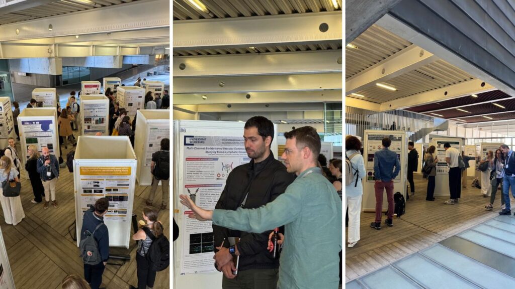
The EMBO Workshop ‘Building networks: engineering in vascular biology’ took place in May at EMBL Barcelona, at the Barcelona Biomedical Research Park (PRBB).
In recent years, advances in multicellular systems, gene editing, stem cell technologies, and bioengineering have created new opportunities to study vascular biology in vitro using human cells. This 2026 installment of #EMBOBuildNetworks reunited researchers working at the intersection of engineering and vascular biology, facilitating discussions on how to apply cutting-edge techniques in spatial omics, advanced imaging, and the use of AI in vascular research.
For this year’s edition of the meeting, we had 150 people attending on-site. There were four financial assistance grants provided by the EMBL Corporate Partnership Programme and EMBO. With a total of 76 posters on display, we held two poster sessions during which presenters could discuss their research. After that, it was time for the speakers to vote and select the winners. There were three best poster prizes awarded during the meeting and we’re pleased to share with you more information on the winners and their research!
How do malaria cues rewire endothelial mechanics?
Authors: Matt Govendir, Waleed Mirza, Adrian Candelas, Juan F. Abenza, Borja Lopez, Martin Bergert, Alejandro Torres Sánchez, Alba Diz-Muñoz, Maria Bernabeu
Presenter: Adrian Candelas

EMBL Barcelona, Spain
Abstract:
Barrier integrity of endothelial tissues relies on a fine balance between intercellular junctional forces and focal adhesion–mediated traction. In cerebral malaria, one of the deadliest complications of Plasmodium falciparum infection, the blood-brain barrier (BBB) is compromised, leading to brain swelling and cerebral haemorrhages that cause over 500.000 deaths annually. Although endothelial disruption is well documented in post-mortem samples, the events that initiate barrier failure remain uncharacterized.
Endothelial barrier function is regulated by mechanosensitive adherens junctions, composed of membrane VE-cadherin homodimers, intracellular protein scaffolds, and a cortical actin network that maintains junctional forces and adherens junction stability.
Combining cutting-edge biophysical microscopy techniques and high-resolution live imaging with mathematical modelling, we show that human brain endothelial cells are rapidly compromised by exposure to P. falciparum products. During the first hours, they transiently accumulate VE-cadherin at junctions, while concurrently recruiting vinculin to focal adhesions. This results in abrupt intercellular junction breakdown, indicative of a fast mechanoresponse to parasite exposure.
After 8 hours, endothelial cells have undergone a profound morphological remodelling, with enhanced numbers of focal adhesions, transversal stress fibre formation, and the acquisition of a migratory phenotype. Traction force microscopy reveals increased matrix forces during this transition, which coincides with elevated FAK activation, a common sign of pathological pro-migratory states.
Altogether, our findings suggest that BBB breakdown upon parasite exposure is driven by an early imbalance between adherens junctions and focal adhesions, leading to mechanical instability, junctional failure, and actin network reorganisation. Importantly, pharmacological inhibition of FAK with PF-573228 preserves endothelial barrier properties, highlighting the importance of endothelial mechanics in cerebral malaria pathogenesis.
Due to the confidentiality of the unpublished data, we cannot share the poster.
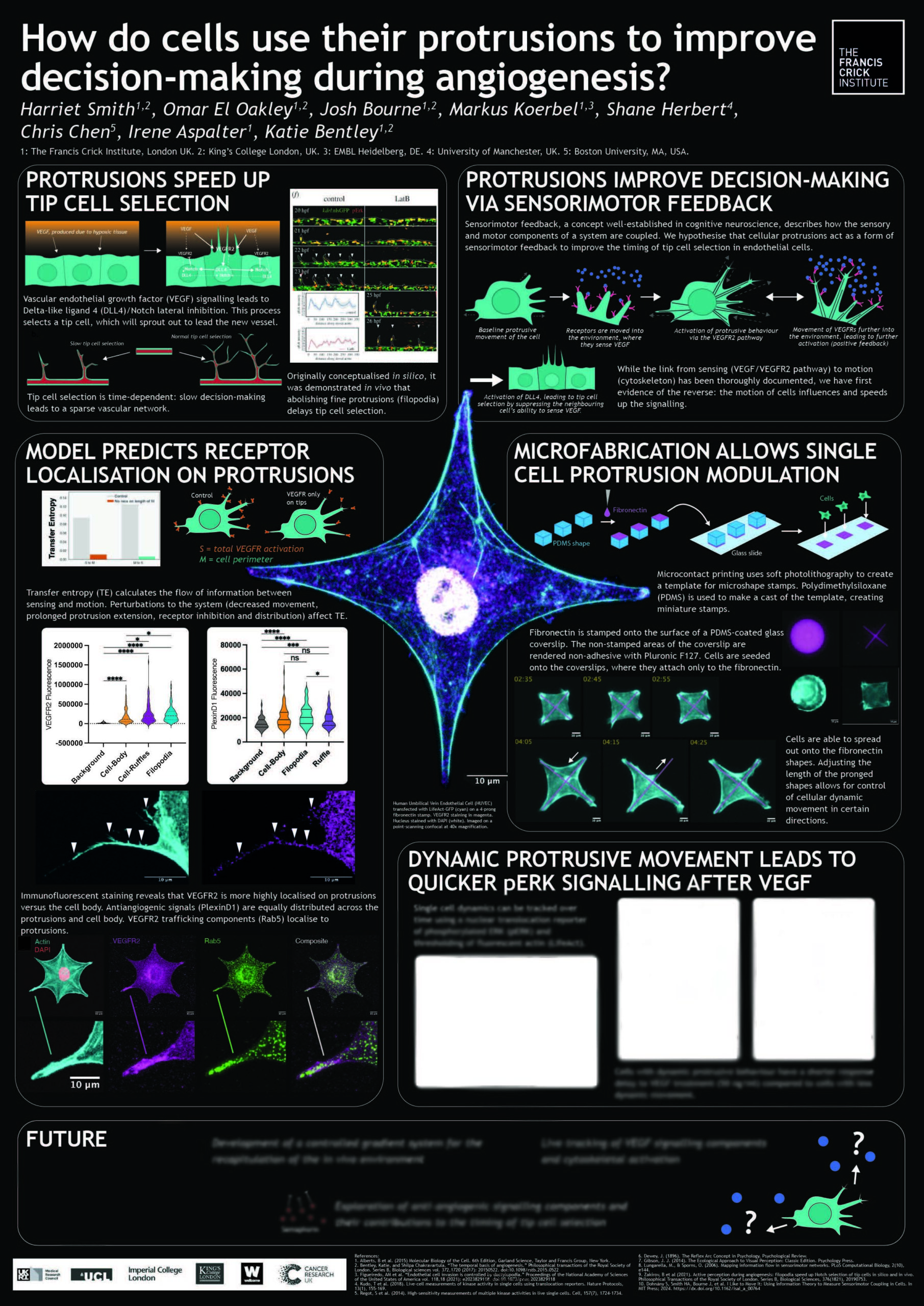
How do cells use their protrusions to improve decision-making during angiogenesis?
Authors: Irene Aspalter, Harriet Smith, Markus Koerbel, Omar El Oakley, Christopher Chen, Katie Bentley
Presenter: Harriet Smith
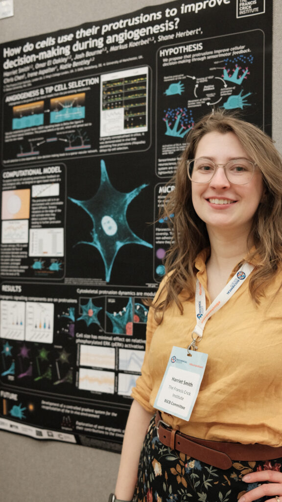
The Francis Crick Institute, UK
Abstract:
Angiogenesis is a complex, dynamic and time-limited process to vascularise and oxygenate tissues. Timely selection of a tip cell to lead the newest sprout towards angiogenic signals is required to keep vascular branching on track. Slower branching decisions can result in sparser network topology – therefore, modulating tip decision timing presents a novel method to tune vascular network topologies in engineered tissues. Previous work from the lab uncovered that dynamic actin-based protrusions on endothelial cells are essential for the timeliness of this decision. Protrusion inhibition leads to slowed tip cell selection and delayed sprouting in silico and in vivo. Here, we integrate microfabricated in vitro and in silico predictive modelling methods to investigate how cells use feedback between movement and sensing (“active perception”) on their protrusions to gather information about the environment to inform collective decision-making: why do protrusions speed up cell decisions? We use an exciting new application of microengineering to explore the tip cell selection process on a single-cell scale. Microfabrication allows us to precisely control cellular dynamic movement, without severely impacting the signalling pathways that would typically be affected by standard cytoskeletal disruptors. We observe that cells with more dynamic movements are quicker to activate downstream (pERK) signalling along the VEGF/VEGFR2 pathway. With the addition of a diffusion-based growth factor gradient, we are able to investigate how context-dependent signalling gradients impact the speed of cell decisions when co-modulating their protrusive ability. Our long-term goal is to leverage our multidisciplinary approach to investigate how cells use protrusive movements to sense both pro and anti-angiogenic pathways as tissue environments in vivo and engineered tissues become more complex. We aim to define the molecular mechanisms impacting these individual cell decision dynamics throughout angiogenesis. The application of these measures is the first step to demonstrating the role of active perception on a cellular level, furthering the field’s understanding of dynamic cell decision-making during tissue construction.
The prize was kindly sponsored by FEBS Letters
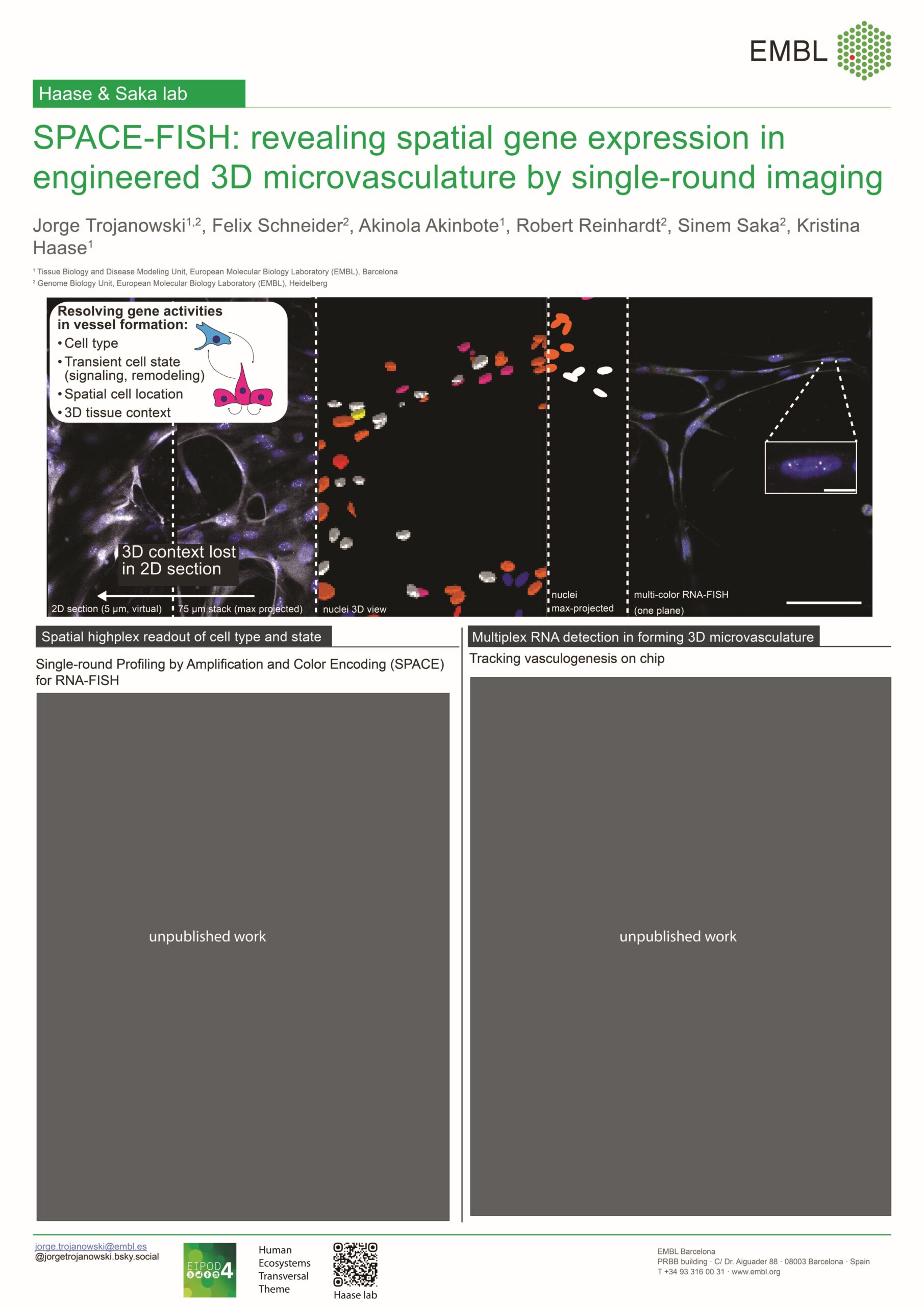
SPACE-FISH reveals spatial gene expression changes in engineered 3D microvascular tissues by multiplexed single round imaging
Authors: Jorge Trojanowski, Felix Schneider, Akinola Akinbote, Robert Reinhardt, Sinem Saka, Kristina Haase
Presenter: Jorge Trojanowski

EMBL Barcelona, Spain
Abstract:
Understanding vascular network formation requires methods that resolve molecular states of individual cells while preserving their three-dimensional spatial organization. Although multiplexed spatial transcriptomics approaches are rapidly advancing, many rely on serial imaging cycles or thin tissue sections, limiting throughput and preventing accurate interrogation of intact 3D tissue organization. Consequently, there remains a need for scalable imaging approaches that enable multiplexed gene expression analysis directly within perfusable 3D vascular models.
Here we introduce Single-round Profiling by Amplification and Color Encoding (SPACE)-FISH, a multiplexed RNA imaging strategy that combines nascent RNA fluorescence in situ hybridization (FISH) with combinatorial color barcoding to profile up to 18 transcripts in a single imaging round. By avoiding iterative staining cycles and enabling imaging in intact samples, SPACE-FISH provides rapid, cost-effective spatial transcript readouts while preserving full 3D positional information.
We applied SPACE-FISH to self-assembled human microvascular networks generated on microfluidic chips, which recapitulate key steps of vasculogenesis and angiogenesis including cell coalescence, sprouting, anastomosis, and lumen formation. Using an angiogenesis-focused gene panel, we mapped transcriptional states across developing vascular structures and identified both global and localized gene expression changes. Notably, we quantified expression gradients along angiogenic sprouts, resolved transcriptionally distinct cellular neighborhoods within vascular plexus regions and in interconnected microvessels, and tracked progressive stromal cell association with forming vessels.
Together, SPACE-FISH establishes a scalable framework for multiplexed gene expression imaging in engineered 3D tissues, enabling systematic investigation of vascular morphogenesis and inflammatory remodeling across experimental conditions, time points, and replicates.
The prize was kindly sponsored by EMBO Press
The EMBO Workshop ‘Building networks: engineering in vascular biology’ took place from 13 – 15 May 2026 at EMBL Barcelona.
Sign up for our newsletter not to miss upcoming events at EMBL Barcelona – and other EMBL sides. Scientists at EMBL Barcelona focus on tissue biology and disease modelling so if you are interested in these areas of research, you can also select the topics you’d like to get updates on from us!