Best poster prizes at ‘Protein synthesis and translational control’
From 3 – 7 September 2025, the EMBL Conference ‘Protein synthesis and translational control’ brought together researchers from around the world to share their latest findings. Sessions covered protein synthesis from the molecular and structural details of the translational machinery to its regulation in cells and organisms, and its role in health and disease. A particular emphasis was placed on mRNA translation, which is key to maintaining cellular homeostasis and responding to environmental, physiological, and pathological signals. The programme highlighted recent advances in translation quality control, new regulators, translation dynamics in living cells, and connections to other cellular processes such as metabolism, as well as implications for diseases including cancer, neurological and metabolic disorders, and viral infections.
#EMBLProtein was attended by 303 participants on site and 71 virtually, with 168 posters displayed at the Advanced Training Centre. From this diverse range of research, six poster prize winners were selected, and we are pleased to introduce them and their work.
Conserved mechanism of collision dependent mRNA degradation
Presenter: Satoshi Hashimoto
Authors: Satoshi Hashimoto, Aoi Satoh, Jiahuan Zeng, Jennifer Sauerland, Hideaki Takeda, Hotaka Kobayashi, Toshifumi Inada

University of Tokyo, Japan
Abstract:
Accurate gene expression is essential for all organisms, but errors during this process are unavoidable. Cells have evolved multiple quality control mechanisms to handle such errors and maintain cellular homeostasis. Among these, No Go Decay (NGD) is a well characterized mRNA surveillance pathway. It prevents the accumulation of problematic mRNAs containing sequences prone to ribosome stalling and collisions, such as tandem rare codons in yeast or poly(A) stretches in mammals.
When a ribosome stalls during translation and collides with a trailing ribosome, a disome is formed (Ikeuchi et al., EMBO J., 2019). In yeast, the disome is specifically recognized by the E3 ubiquitin ligase Hel2, which polyubiquitinates the ribosomal protein uS10, triggering ribosome associated quality control (RQC) to degrade the aberrant nascent polypeptide (Matsuo et al., Nat. Commun., 2017; NSMB, 2020). Ribosome collision also activates No Go Decay, and Cue2 is responsible for the endonucleolytic cleavage at the vicinity of the collided ribosome (D’Orazio et al., eLife, 2019), depending on the Hel2 mediated polyubiquitination of uS10 and eS7 (Ikeuchi et al., EMBO J., 2019; Tomomatsu et al., NAR, 2022). In addition, Syh1 is proposed to promote mRNA decay upon ribosome stalling by recruiting the exonuclease Xrn1, thereby triggering the exonuclease mediated decay pathway (Veltri et al., eLife, 2022). However, it remains unclear how both pathways, Hel2 Cue2 dependent endonucleolytic cleavage, and Syh1 Xrn1 dependent exonucleolytic degradation, contribute to the ribosome collision mediated mRNA decay.
Here, we demonstrate that in yeast, Syh1 Xrn1 and Hel2 Cue2 pathways independently function in the ribosome collision induced mRNA decay. In addition, we confirm that these mechanisms are conserved in mammals by using a Tet Off system and single molecule imaging. These findings deepen our understanding of mRNA quality control and offer the potential for developing mRNA targeted therapies.
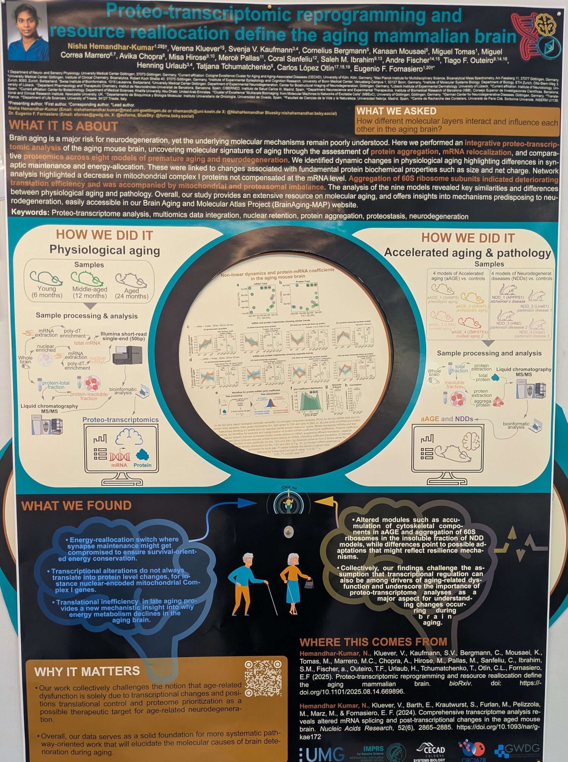
Proteo-transcriptomic reprogramming and resource reallocation define the aging mammalian brain
Presenter: Nisha Hemandhar Kumar
Authors: Nisha Hemandhar Kumar, Eugenio Fornasiero, Verena Kluever, Svenja Kaufmann, Carlos Lopez Otin, Tatjana Tchumatchenko, Henning Urlaub, Tiago Outeiro, Andre Fischer, Saleh M. Ibrahim, Mercè Pallàs, Misa Hirose, Miguel Correa Marrero, Kanaan Mousaei, Cornelius Bergmann

University of Cologne, Germany
Abstract:
Brain aging is a major risk for neurodegeneration, yet the underlying proteo transcriptomic mechanisms remain poorly understood. Here we performed an integrative proteo transcriptomic analysis of the aging mouse brain, uncovering molecular signatures of aging through the assessment of protein aggregation, mRNA relocalization, and comparative proteomics across eight models of premature aging and neurodegeneration. We identified dynamic changes in physiological aging highlighting differences in synaptic maintenance and energy allocation. These were linked to changes associated with fundamental biochemical properties such as protein size and net charge. Network analysis highlighted a decrease in mitochondrial complex I proteins not compensated at the mRNA level. Aggregation of 60S ribosome components indicated deteriorating translational efficiency leading to mitochondrial and proteasomal imbalance. The analysis of the nine models revealed key similarities and differences between physiological aging and pathology. Overall, our study provides an extensive resource on molecular aging, and offers insights into mechanisms predisposing to neurodegeneration, easily accessible soon after publishing.
View spinning poster video and view image of spinning poster
Multi-scale analysis of mRNA translation adaptation to nutrient shifts reveals novel regulatory elements
Presenter: Xavier Hernandez Alias
Authors: Xavier Hernandez Alias, Selay Kaya, Julien Gagneur, Danny Nedialkova
Max Planck Institute of Biochemistry / Technical University of Munich, Germany
Abstract:
Cells react to environmental stimuli by modulating gene expression across different levels and time scales. To investigate fast and dynamic changes in mRNA translation, we conducted simultaneous time resolved measurements of changes in mRNA abundance and ribosome loading in budding yeast subjected to three distinct shifts in nutrient availability. Using spike in calibrated ribosome profiling, we found that translational regulation occurs within minutes and is much faster than changes in mRNA abundance. Notably, upon glucose upshift, 66% of mRNAs exhibited altered ribosome occupancy within the first 10 minutes, compared to only 22% showing changes in mRNA levels. In contrast, adaptation to nitrogen upshift primarily involved mRNA abundance regulation with translational potentiation, while phosphate upshift triggered a highly transient translational response, specifically targeting to phospho related gene sets. To uncover the underlying regulatory code, we developed RiboNet, a deep learning model that predicts absolute ribosome counts and their distribution along endogenous mRNA transcripts. RiboNet predicts ribosome occupancies that achieve or surpass experimental replicate accuracy and accounts for most of the variance observed across time resolved analysis of nutrient shift responses. Designed as a multi task framework, RiboNet enables flexible model interpretation through in silico mutagenesis of regulatory sequences and comparisons between conditions. Our analysis identified coding sequences as the major determinants of ribosome occupancy, uncovering differential and transient elongation pauses at specific codons upon glucose and nitrogen shifts, as well as novel regulatory motifs in 5’ UTRs. Our multi scale approach combining modeling and experimental data thus reveals previously unrecognized regulatory layers of translational control in the yeast stress response.
Due to the confidentiality of the unpublished data, we cannot publish the poster.
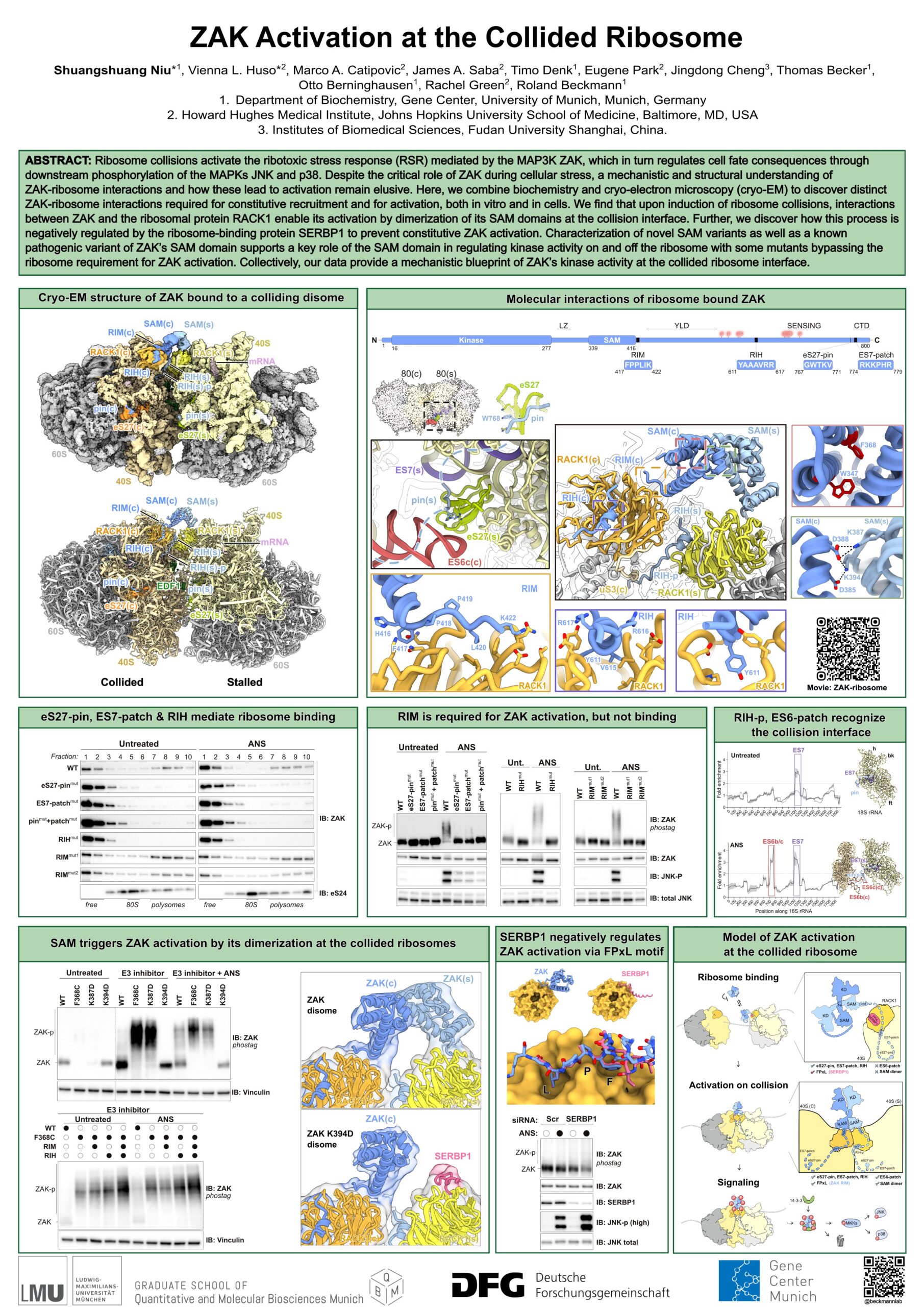
Structural decoding of ribosome collision sensing by ZAKa
Presenter: Shuangshuang Niu
Authors: Timo Denk, Vienna Huso, Shuangshuang Niu, James Saba, Eugene Park, Marco Catipovic, Jingdong Cheng, Thomas Becker, Rachel Green, Roland Beckmann
Ludwig Maximilians University of Munich, Germany
Abstract:
The MAP3K ZAKα plays a key role in sensing persistent ribosome collisions and acts as the initiator of the ribotoxic stress response (RSR). Its activation triggers downstream phosphorylation of stress activated MAP kinase p38 and Jun N terminal kinase JNK. Despite its critical role in determining cell fate, ranging from cell cycle arrest to cell death, the structural and mechanistic basis underlying ZAKα activation upon ribosome collision has remained elusive.
In this work, we present the cryo electron microscopy structures of ZAKα bound to ribosomes in normal condition and stress induced condition. We demonstrate that the collision sensing capabilities of ZAKα depend on multiple distinct regions, and we further assess their respective impacts on ribosome binding and ZAKα activation. These findings lead to a mechanistic model explaining how ZAKα is activated at the collided ribosome.
Leaderless mRNAs in bacteria are well-translated and regulated by distal elements
Author and presenter: Juliana Stanley
Massachusetts Institute of Technology, United States of America
Abstract:
Many bacterial mRNAs lack the 5′ untranslated region that is considered key to recruiting ribosomes. How these “leaderless mRNAs” (lmRNAs) initiate translation remains unknown. Here we show that lmRNAs native to E. coli are translated as efficiently as canonical leadered mRNAs in vivo and in vitro, and that this efficiency requires specific sequences located >150 bases downstream of the start codon. In contrast to native lmRNAs, translation is dramatically reduced for artificial lmRNAs consisting of either heterologous or native genes with their leaders removed. Translation of these artificial lmRNAs can be rescued by transplanting specific regions of native lmRNAs in vivo. Our results indicate that lmRNAs use downstream sequence to mediate translation, potentially by recruiting ribosomes or additional factors. This work furthers our understanding of the map between RNA sequence and expression level. It also lays the groundwork for investigating the regulation of protein synthesis in many medically important bacteria, such as Mycobacterium tuberculosis, whose mRNAs are often leaderless.
Due to the confidentiality of the unpublished data, we cannot publish the poster.
Poster prize kindly sponsored by FEBS Letters
Calmodulin as an essential chaperone for co-translational folding of the Kv7.2 ion channel
Presenter: Jack Tait
Authors: Jack Tait, Arantza Muguruza Montero, Sander Tans, Ane Metola, Gunnar von Heijne, Vanda Sunderlikova, Alexandros Katranidis, Eider Nuñez, Sara M Alicante, Alvaro Villarroel

AMOLF, The Netherlands
Abstract:
In vivo, a majority of nascent protein chains must begin folding during translation in order to obtain their native structure. While the importance of co translational folding has become increasingly clear, data on the specific mechanisms underlying the necessary coordination between the ribosome, nascent chain and molecular chaperones during co translational folding have proven challenging to obtain. Here, we construct a complete model of the co translational folding pathway of the calcium responsive domain (CRD) of the Kv7.2 human neuronal ion channel, and demonstrate that interactions with calmodulin (CaM) are crucial for native folding of the domain. By combining force profile analysis with ensemble FRET and single molecule force spectroscopy techniques, we identify the key folding stages of the CRD during translation, determining the conformation of the nascent chain at each stage. We show that binding of CaM at each stage induces the formation of a metastable hairpin, stabilising the nascent CRD against formation of off pathway misfolds until the full domain has been translated. These findings expand on the role of CaM as a key regulator of folding in eukaryotes: not only as an essential cellular signalling protein, but also as a bona fide molecular chaperone.
The EMBL Conference ‘Protein synthesis and translational control‘ took place from 3 – 7 September 2025 at EMBL Heidelberg and virtually.