Meet the poster prize winners of ‘The complex life of RNA’
The EMBO | EMBL Symposium ‘The complex life of RNA’ brought together 425 scientists at EMBL Heidelberg from 13 – 16 October 2026 to discuss the latest advances in RNA biology. Covering topics ranging from co-transcriptional RNA biology and RNA processing to RNA structure, regulation and therapeutics, the symposium explored the many roles RNA plays in the transmission and regulation of genetic information across all forms of life. 25 financial assistance grants were awarded through the EMBL Corporate Partnership Programme and EMBO.
With 275 posters on display, participants had plenty of opportunities to share their research and exchange ideas with colleagues across the field. Out of these various posters, nine winners were chosen by the participants. We are delighted to congratulate them on their excellent work and share the research of seven of them!
Detection of putative bifunctional genes through genome-wide long-read RNA sequencing and polysome profiling
Authors: Hansoll Na, Ngoc Bao To, Kyung-Tae Lee, Jin-Wu Nam
Presenter: Ngoc Bao To

Hanyang University, Republic of Korea
Abstract:
Background: The traditional distinction between protein-coding and non-coding genes is increasingly challenged by evidence that a single gene can generate both coding and non-coding transcript isoforms. The extent, mechanisms, and functional significance of such bifunctional genes remain poorly understood.
Methods: To systematically identify bifunctional genes, we combined long-read (LR) and short-read (SR) RNA sequencing with polysome profiling in normal colonic epithelial (CCD841) and colorectal cancer (DLD1) cell lines. Long-read sequencing enabled full-length transcript characterization, allowing accurate discrimination of coding and non-coding isoforms in unbound and heavy polysome fractions. A bifunctionality score was developed to quantify the dual coding and non-coding potential of each gene, which is computed by representing each transcript as a vector of its heavy-polysome and unbound expression and taking the maximum cross-product magnitude over all coding and non-coding transcript pairs.
Results: We identified 764 bifunctional genes in CCD841 cells and 556 in DLD1 cells, with consistent detection across polysome fractions. Shared bifunctional genes were enriched in ion transport, calcium signaling, and TNFα-related pathways, while cell line-specific genes were associated with mitochondrial metabolism in CCD841 and chromatin organization and developmental processes in DLD1. Alternative splicing was the primary mechanism underlying bifunctionality, accounting for over 75% of identified genes. Importantly, isoform-specific qPCR validated the co-expression of coding and non-coding transcripts from selected candidates, supporting the accuracy of the bifunctionality scoring framework.
Conclusions: Our findings demonstrate that bifunctional genes are widespread and predominantly generated through alternative splicing. Integrating isoform-resolved transcriptomics with experimental validation establishes bifunctionality as an important layer of gene regulation and highlights its potential role in cancer development and therapeutic targeting.
Due to the confidentiality of the unpublished data, we cannot share the poster.
The prize was kindly sponsored by KeAI Publishing.
The periphery of nuclear speckles defines a spatially and temporally regulated compartment of long-lived intron-retained RNAs that resolves during mitosis
Authors: Josep Biayna Rodríguez, Artem Baranovskii, Anusha Chaudhuri, Marta Paladin , Berkay Erdem, Luise-Elektra Keller, A Rasim Barutcu, Stefanie Dimmeler, Annalisa Marsico, Gabrijela Dumbović
Presenters: Josep Biayna Rodríguez and Marta Paladin
Goethe University Frankfurt, Germany
Abstract:
RNA localization adds a fundamental layer to gene expression by determining when and where translation-ready mRNAs become available. Here, we identify a subnuclear RNA niche at the nuclear speckle periphery that couples intron retention to cell-cycle-timed RNA release. Using compartment-resolved transcriptional inhibition, sequence-based deep learning, and single-molecule and super-resolution RNA imaging in human pluripotent stem cells, we define a class of nuclear RNAs with long-lived retained introns (LL-IR-RNAs) that maintain stable intron retention for hours and are enriched in genome maintenance and mitosis regulators, including centromere and kinetochore assembly, DNA repair, and telomere maintenance. Long-lived retained introns are characterized by elevated GC content, predicted structural stability, and association with nuclear speckle RBPs. In interphase, LL-IR-RNAs localize to a distinct nuclear speckle-peripheral niche conserved across cell types. During mitotic remodeling, they undergo coordinated, kinase-dependent splicing and populate the cytoplasm of early G1 daughter cells. These findings link cis-encoded intronic features, subnuclear organization and mitotic remodeling to temporal control of RNA fate.
The prize was kindly sponsored by the RNA Society.
Regulation of human HBB expression by non-canonical translation in the 3′ untranslated region
Authors: Japita Ghosh, Kritika Chauhan, Debaleena Kar, Sandeep M Eswarappa
Presenter: Japita Ghosh

Indian Institute of Science, India
Abstract:
Regulation of gene expression is crucial for proper cellular function. In human mature erythrocytes, post-transcriptional control is the only mechanism of gene expression regulation, as they are anucleate and transcriptionally inactive. The erythrocyte proteome consists mainly of haemoglobin protein, a tetramer of alpha- (HBA) and beta-globin (HBB). So, HBA and HBB expression must be tightly regulated to maintain the balance of globins and thus erythrocyte function. Here, we studied the regulation of human HBB expression, mediated by non-canonical translation in its 3′ untranslated region (UTR). We first demonstrated that HBB mRNA undergoes stop codon readthrough (SCR), a process in which translation is continued beyond the canonical stop codon to generate a C-terminally extended protein isoform. Thus, SCR in the HBB transcript produces a unique proteoform, the beta-globin readthrough protein with a 20-amino-acid C-terminal extension, along with the canonical protein. Analysis of available mass spectrometry data from human erythrocytes also confirmed the presence of peptides from the C-terminal extension. Further investigations into the functional significance of SCR indicated that the beta-globin readthrough protein is unstable and its expression is rescued by proteasomal inhibitors, indicating that the readthrough peptide is prone to proteasomal degradation. All these data suggest that SCR negatively regulates canonical beta-globin expression. Later, in silico analysis of publicly available ribosome profiling data demonstrated dense ribosomal footprints in the 3′UTR of HBB beyond the SCR-undergoing region. Further analysis indicated that the readscorrespond to a putative small downstream ORF (dORF), potentially encoding a 10-amino-acid-long micropeptide. Experiments revealed that the HBB dORF gets translated in a cap-dependent manner. Further studies confirmed that disrupting the translation of the HBB dORF impairs expression of the main ORF, indicating a positive regulation of the dORF translation on the main ORF expression. Thus, our results reveal that two distinct non-canonical translation events in the 3′UTR of the HBB transcript act antagonistically to regulate HBB expression.
Due to the confidentiality of the unpublished data, we cannot share the poster.
The prize was kindly sponsored by Oxford University Press.
A two-step chromosome-retention mechanism enables pre-rRNA inheritance through mitosis
Authors: Zuzana Koskova, Alberto Hernandez-Armendariz, Yuki Hayashi, Ina Huppertz, Sara Cuylen-Häring
Presenter: Zuzana Koskova

EMBL Heidelberg, Germany
Abstract:
Pre-ribosomal RNAs (pre-rRNAs) are large, highly structured scaffolds for pre-ribosome assembly inside of the nucleolus. During mitosis, nucleolar disassembly releases these RNAs into the cytoplasm, raising the question: can the pre-rRNAs be re-incorporated into the nascent nucleus and enter a new cycle of ribosome biogenesis? Previous studies have demonstrated that pre-rRNAs and nucleolar proteins are recruited to the surface of mitotic chromosomes by the intrinsically disordered protein Ki-67, but the mechanism by which Ki-67 selectively enriches nucleolar material and the functional relevance of this chromosomal enrichment remained unclear.
Here, we show that the selective enrichment of nucleolar material on chromosomes is mediated by a small canonical pre-rRNA-binding protein, which links Ki-67 to pre-rRNAs in early mitosis in a phosphorylation-dependent manner. Depletion of this adapter protein causes pre-rRNAs as well as nucleolar proteins to localize to large cytoplasmic aggregates, as observed upon Ki-67 depletion. During mitotic exit, though, a widespread dephosphorylation uncouples the adapter protein with bound pre-rRNAs from Ki-67. Instead, the RNAs are retained on chromosomes by a distinct Ki-67 domain through a phase separation mechanism. Loss of this Ki-67 domain weakens the pre-rRNA anchoring, allowing the reforming nuclear envelope to displace pre-rRNAs and associated factors from the surface of mitotic chromosomes into the cytoplasm, thereby excluding them from the newly formed nucleus and resulting in smaller nucleoli. Thus, this two-step process of selective enrichment followed by phase separation preserves nucleolar material on chromosomes and enables rapid nucleolar reassembly after cell division.
Overall, we trace the journey of pre-rRNAs through mitosis and demonstrate a new principle of cellular organization: pre-rRNAs are pre-compartmentalized on mitotic chromosomes, ensuring immediate nuclear and nucleolar incorporation after cell division.
Due to the confidentiality of the unpublished data, we cannot share the poster.
The prize was kindly sponsored by Febs Letters.
Composite SMG5-SMG6 PIN domain formation is essential for human nonsense-mediated mRNA decay
Authors: Katharina Kurscheidt, Sophie Theunissen, Natalia Pasquali, Kerstin Becker, Volker Boehm, Elena Conti, Niels H. Gehring
Presenter: Katharina Kurscheidt

Max Planck Institute of Biochemistry, Germany
Abstract:
The nonsense-mediated mRNA decay (NMD) pathway relies on the coordinated assembly and action of multiple protein factors for the recognition and elimination of transcripts harbouring premature termination codons (PTCs).
The degradation of targets is initiated by an endonucleolytic cleavage event near the PTC mediated by the SMG6 endonuclease and requires the presence of the downstream effector SMG5. Both SMG6 and SMG5 contain C-terminal PIN domains. While the SMG6 PIN domain contains the catalytic tetrad of carboxylate residues and accordingly exhibits weak nucleolytic activity in vitro, the SMG5 PIN domain lacks the catalytic residues and is inactive. However, the molecular details of endonuclease regulation and activation as well as the mechanistic role of SMG5 in SMG6-mediated cleavage have remained elusive.
Combining structural predictions with in vitro nuclease assays, and NMD rescue assays in cultured cells, we show that SMG5 and SMG6 interact via their PIN domains to form a composite interface (cPIN) with functional relevance. In vitro reconstituted SMG5-SMG6 cPIN heterodimers show high activity, as SMG5 completes the SMG6 active site and contributes to substrate binding. Mutations targeting the predicted PIN-PIN interface, the RNA-binding site, or the catalytic centre abolish the SMG5-stimulated SMG6 activity in vitro and inhibit cellular NMD.
In conclusion, our findings demonstrate how paralogous PIN domains complement each other to assemble a highly active endonuclease in NMD, providing a structural and mechanistic explanation for efficient NMD substrate degradation.
The prize was kindly sponsored by KeAI Publishing.
4TU-Live-seq reveals coupling of transcription elongation and post-transcriptional splicing in living vertebrates
Authors: Markus Vogt, Ashwin Narain, Rahaf, Issa, Nevenka Dudvarski Stankovic, Pranjali Bhandare, Yiliam Cruz Garcia, Teresa Rummels, Anton Hamann, Daniel Baumann, Stefan Knapp, Florian Erhard, Elmar Wolf
Presenter: Markus Vogt
University of Kiel, Germany
Abstract:
Pre-mRNA splicing is central to shaping gene expression and generating proteome diversity. Although transcription and splicing both take place in the nucleus, the degree to which splicing occurs co-transcriptionally in living multicellular organisms, and whether ongoing transcription is required for its efficiency, has remained poorly defined. Splicing kinetics have so far been characterized almost exclusively in cell extracts or cultured cell lines, leaving splicing dynamics in living vertebrates largely unexplored due to technical constraints.
To close this gap, we established 4TU-Live-seq, a metabolic labeling method built on 4-thiouracil (4TU) incorporation followed by its chemical conversion into a cytosine analog. Applying this approach to a pancreatic cancer allograft model, we labeled nascent transcripts in engrafted tumor cells over 30–120 minute windows, derived unbiased splicing ratios, and fit a first-order kinetic model to obtain absolute intron-level splicing rates.
Splicing proceeded with a median time of roughly 6 minutes, though individual introns spanned a broad range from 2 to over 30 minutes. To identify the determinants of this variability, we trained a machine-learning classifier to separate fast- from slow-spliced introns using architectural features (intron length, position) together with cis-regulatory sequence elements. Polypyrimidine tract strength emerged as the strongest positive predictor of splicing speed, while high GC content at the 3′ splice site was most strongly associated with slower splicing.
Based on these kinetic estimates, most vertebrate introns are resolved post-transcriptionally. Nonetheless, acutely depleting the elongation factor SPT6 via auxin-induced degradation impaired splicing efficiency, demonstrating that productive transcription elongation remains necessary for optimal splicing outcomes in vivo.
Lastly, genes with the fastest in vivo splicing kinetics were enriched for pathways underlying cellular adaptation to organismal physiology, pointing to splicing speed as an evolutionarily tuned, rate-limiting layer of regulation for specific gene expression programs.
Due to the confidentiality of the unpublished data, we cannot share the poster.
The prize was kindly sponsored by Oxford University Press.
Mapping Sm Protein Binding Sites on snRNA Using RNP-MaP
Authors: Jarrett Wilson, Chase Weidmann
Presenter: Jarrett Wilson

University of Michigan, USA
Abstract:
RNA-protein interactions influence every step of gene regulation, governing RNA transcription, splicing, localization, stability, and translation and ultimately shape cell identity and fate. However, defining the organization and function of RNA-protein interaction networks remains a major challenge, particularly because many RNAs lack strong primary sequence conservation or well-defined structural annotation. Traditional approaches to study RNA-protein interactions, for example, RNA immunoprecipitation or reconstituted binding assays, often miss the dynamic and cooperative nature of Ribonucleoprotein (RNP) assemblies inside cells and are limited to the interactions of only a few proteins at a time. In contrast, RNP-MaP (Ribonucleoprotein Networks Analyzed by Mutational Profiling) comprehensively maps the locations of protein binding sites on RNA and measures interactions between RNA binding proteins on single RNA molecules inside cells. Here, we aim to identify RNP networks transcriptome-wide based on their RNP-MaP interaction network signatures. As proof-of-principle, we are investigating interactions between the Sm-protein complex and small nuclear RNAs (snRNAs), which together are central to spliceosomal function. Sm proteins assemble at a conserved Sm binding site, forming well-defined complexes that provide an ideal system for characterizing RNP network formation. Our data reveals consistent RNP-MaP reactivity patterns at the Sm binding site across multiple snRNAs. From these shared reactivity profiles, we constructed a machine learning-based model that accurately identifies Sm binding events from small, targeted RNP-MaP datasets. We aim to use our model to identify novel Sm binding sites from transcriptome-scale RNP-MaP data. This approach will serve as a framework for identifying new RNP-MaP signatures and discovery of novel RNP networks inside the cell.
The prize was kindly sponsored by KeAI Publishing.
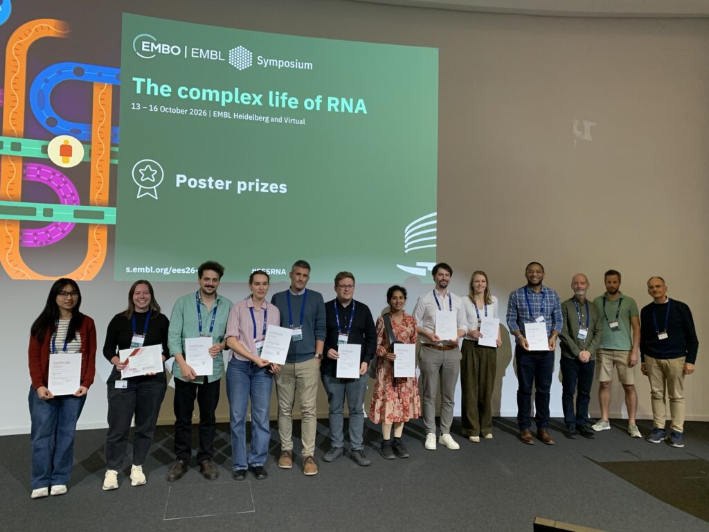
The EMBO | EMBL Symposium ‘The complex life of RNA‘ took place from 13 – 16 October 2026 at EMBL Heidelberg and virtually.
Find out more about the #EESRNA meeting from the blog post written by Luciano Marasco, who participated as an event reporter.