Meet the best poster prize winners of ‘Mechanobiology across the tree of life’
From 9 – 12 June 2026, we welcomed 160 on-site and 49 virtual participants to the EMBO | EMBL Symposium ‘Mechanobiology across the tree of life‘, bringing together researchers from diverse biological systems.
The symposium explored the latest technologies and approaches advancing our understanding of mechanobiology, from cell walls and cytoskeletons to membrane biology. Discussions covered advanced imaging techniques in in vitro and in vivo systems, alongside molecular biology, computational methods, and biophysics approaches. With a strong focus on early-career researchers, the meeting aimed to encourage knowledge sharing and foster collaborations across research communities and biological kingdoms.
Participants also shared their latest findings through 98 poster presentations, showcasing the breadth of research in the field. Five presenters received poster prizes for their outstanding contributions.
Read on to discover more about their research and join us in congratulating this year’s winners!
Design and self-assembly of cytomotive filaments
Presenter: Marija Krstic
Authors: Marija Krstic, Maitane Muñoz-Basagoiti, Christian Vanhille-Campos, Andela Šaric, Buzz Baum

Institute of Science and Technology Austria, Austria
Abstract:
Many cytoskeletal filaments that drive cellular shape changes and motility, such as actin and FtsZ, undergo treadmilling—a dynamic process in which monomers continuously add to one filament end while dissociating from the other. However, the physical principles that enable treadmilling remain unclear.
Treadmilling requires monomer structure to encode faster binding at one filament end to establish directionality. Hydrolysis, modelled as stochastic bond weakening, promotes depolymerisation at the opposite end. Because hydrolysis generates a gradient of weakened monomers, filaments must remain stable in the bulk while disassembling at the ends to avoid fragmentation. How single-stranded filaments (e.g., FtsZ) satisfy these constraints is unknown.
To probe the single-filament requirements for treadmilling, we construct a colloidal monomer that polymerises into single-stranded filaments and systematically identify the design features required for sustained treadmilling.
We first investigate how directionality can be encoded at the monomer level by analysing binding to a non-hydrolysing filament whose ends are exposed to monomer pools at constant concentration. We find that a way to encode binding kinetics asymmetry is through geometrically modifying the accessible binding area via a conformational switch, leading to asymmetric growth rates.
We then incorporate hydrolysis by stochastically weakening binding interfaces within the filament. As monomer–monomer binding strength decreases, analysis of filament stability shows that a two-state system is necessary to stabilise monomers in the filament interior while permitting end disassembly.
Combining these ingredients yields a monomer architecture that exhibits sustained treadmilling dynamics. These results establish general design principles for engineering treadmilling polymers from simple components.
Kindly sponsored by The FEBS Journal.
Decoding the mechanical state of cells and tissues: an inverse statistical
Presenter: Mathis Grelier
Authors: Mathis Grelier, Maja Milas, Madhura Ramani, Ana-Suncana Smith, Mark Schvartzman, Carlos Ureña Martin

Friedrich Alexander Universität (FAU), Germany
Abstract:
Quantifying mechanical properties-such as cortical tension, adhesion energy, and elasticity- typically relies on invasive techniques like AFM or laser ablation. In contrast, cell morphology itself can be used as a physical readout of these internal forces. We propose a non-invasive, data-driven alternative: extracting these physical parameters directly from static microscopy images using an inverse statistical mechanics framework.
Our approach treats shape variability as steady-state fluctuations. By defining an effective energy functional acting on an appropriate configurational space of shapes, and weighing the probability of observing specific morphologies, we can infer the hidden mechanical parameters associated with a particular steady state. Through a multi-parametric optimization procedure, the model can be fitted to experiments, yielding all mechanical parameters at once. We first validated this method using single cells on substrates with micro-patterned stiffness heterogeneities. We successfully reconstructed the mechanical state of cells undergoing stiffness-induced contact guidance, disentangling the energetic contributions of cell elasticity, cortical tension, and substrate adhesion. These predictions were quantitatively verified against pharmacological perturbations of the cytoskeleton.
Building on this validation, we extend the framework to analyze confluent tissues on uniform substrates. We adapt the effective energy functional to account for intercellular coupling and topology, allowing us to interpret the shape statistics of individual cells within a monolayer. This establishes a high-throughput computational tool capable of mapping mechanical heterogeneity in tissues solely from geometrical data, circumventing the need for physical contact or complex experimental setups.
Kindly sponsored by The FEBS Journal.
Ensnarled: Deciphering the architecture and physiology of complex interconnected networks
Presenter: Chinmayi Subramanya
Authors: Chinmayi Subramanya, Yu Tian, Carl Modes

Max Planck Institute of Molecular Cell Biology and Genetics, Germany
Abstract:
In the vast realm of complex systems, the interplay between multiple networks is a recurring motif that permeates various domains, from crystal lattices and polymers to vascular systems and supercoiled DNA structures. Many biological systems organize through interacting filamentous and membranous networks whose architecture both constrains and enables mechanical function. We introduce a quantitative framework to characterize structural interlinking between networks, which we term ‘ensnarlment’. The approach combines spatial network reconstruction with cyclic representations and Gauss linking numbers, enabling the construction of a novel ensnarlment operator whose spectral properties identify structurally critical elements that organize global architecture. Rather than focusing on a single system, we show that ensnarlment provides a unifying description across biological organization levels, capturing how such systems emerge during growth and self-organization and how topological organization changes during adaptation or pathological remodeling. This work establishes a novel framework for analyzing and characterizing the structural properties of intertwined networks, and provides a foundation for future investigations into the intricate nature of organ functionality with potential implications for organ development, disease, and regeneration.
Due to the confidentiality of the unpublished data, we cannot show the poster.
Uncoupling adhesion- and density-driven rigidity transitions triggers epithelial organisation in embryonic tissues
Presenter: Laura Rustarazo-Calvo
Authors: Laura Rustarazo-Calvo, Elisa Floris, Cristina Pallares, Maximilian Hingerl, Adrian Aguirre Tamaral, Camilla Autorino, Arif ul Maula Khan, Bernat Corominas-Murtra, Nicoletta Petridou
EMBL Heidelberg, Germany
Abstract:
The active regulation of tissue material properties via phase transitions is central in morphogenesis. Transitions abruptly occur at critical points in diverse control parameters, including cell density, shape or adhesion. Whether these parameters are interdependent, performing redundant or distinct functions, is unknown. Here we show that depending on the co-regulation of multiple control parameters, a tissue not only tunes its deformability, but also its morphogenetic trajectory. We theoretically define a new phase diagram capturing the material states of zebrafish pluripotent tissues undergoing epiboly and show that they cross simultaneously critical points in cell density, connectivity and adhesion strength. Combining optogenetics, biophysical measurements, and quantitative morphometrics, we independently modulate each parameter in vivo, identifying adhesion as the main determinant of tissue rheology. Unexpectedly, uncoupling adhesion from density and inducing adhesion-driven rigidification in unjammed pluripotent tissues is sufficient to switch their morphogenetic program and trigger epithelial organisation. This switch in tissue reorganisation is achieved via tricellular junction formation, followed by lumenogenesis and initiation of apical polarity. Altogether, this work reveals the non-linear dynamics of emergent tissue mechanics as instructive mechanisms of tissue organisation and morphogenesis.
Mechanical fracturing of the extracellular matrix patterns the vertebrate heart
Presenter: Daniel Santos-Olivan
Authors: Daniel Santos-Olivan, Alejandro Torres Sánchez, Christopher Chan Jin Jie, Rashmi Priya
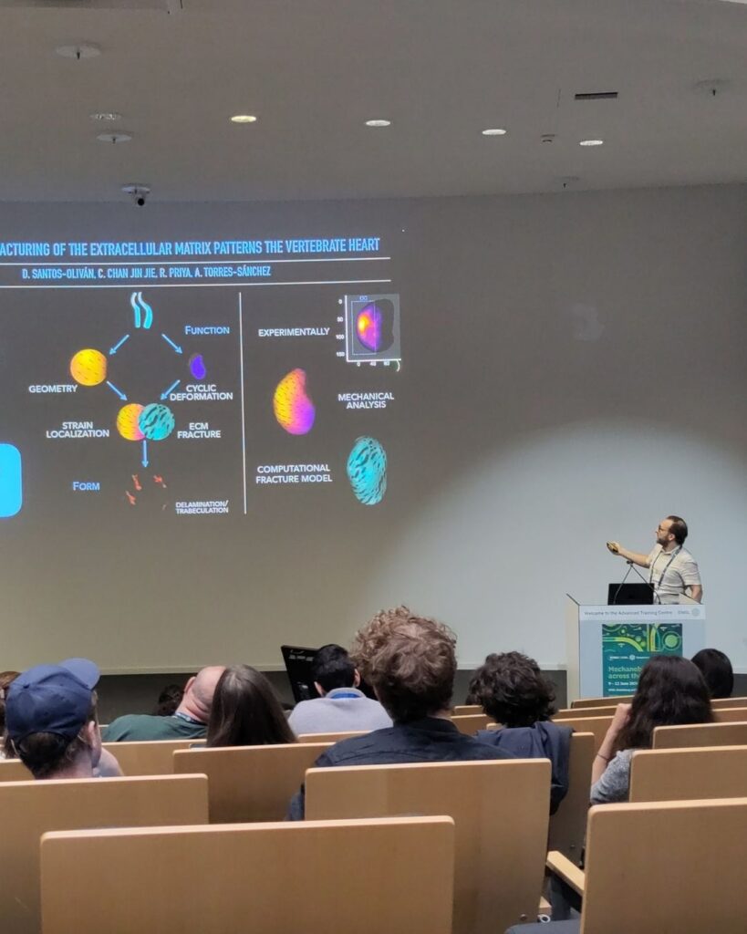
EMBL Barcelona, Spain
Abstract:
Trabeculation, the process where cardiomyocytes delaminate to form ventricular ridges essential for circulation, is a critical step during heart development. While previously attributed only to biochemical prepatterning, we demonstrate that trabeculation is instead guided by mechanical fracturing of the cardiac extracellular matrix (cECM), induced by myocardial contractions. The heart’s anisotropic curvature directs mechanical stress, leading to spatially patterned cECM fractures that serve as structural cues for trabeculation. To investigate this mechanism, we quantify the strain produced by the myocardial layer and develop a biophysical mathematical model that simulates cECM dynamics under cyclic cardiac beating. Using computational simulations on a 3D reconstruction of the myocardium, we predict fracture patterns based on tissue geometry and material properties, matching experimental observations in zebrafish embryos. These findings establish mechanical fracturing as a direct morphogenetic driver of trabeculation, challenging the paradigm that biochemical signals alone dictate ventricular patterning. More broadly, our work underscores how tissue geometry and mechanical forces actively shape cardiac development, reinforcing a self-organizing principle in morphogenesis.
Due to the confidentiality of the unpublished data, we cannot show the poster.
The EMBL Conference ‘Mechanobiology across the tree of life‘ took place from 9 – 12 June 2026 at EMBL Heidelberg and virtually.