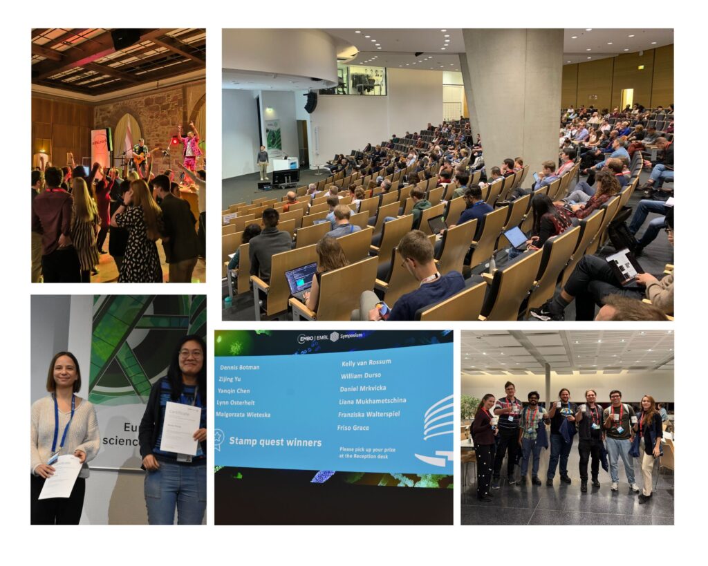
Meet the poster prizes winners of ‘Seeing is believing: imaging the molecular processes of life’
The EMBO | EMBL Symposium ‘Seeing is believing: imaging the molecular processes of life’ took place three weeks ago at EMBL Heidelberg and virtually.
This long-standing symposium has been know to embrace novel imaging technologies that open new windows for biological discovery, including single-molecule and super-resolution, light sheet, and correlative light electron microscopy.
During the four days of the conference researchers from diverse backgrounds and leading developers of imaging methods with cutting-edge applications came together for presentations, discussions, and interactions between students, postdocs, junior and senior investigators.
We welcomed 326 on-site and 114 virtual participants. 17 fellowships were awarded through the EMBL Corporate Partnership Programme and EMBO.
Special for this symposium was that there was a poster prize committee that voted for their favourites among the 125 posters presented.
Congratulations to the three winners! We are pleased to share their abstracts below.
Event-driven microscopy with real-time perturbation to investigate mitochondrial network dynamics
Presenter: Alara Kiris
Authors: Alara Kiris, Giorgio Tortarolo, Willi Stepp, Juan Cruz Landoni, Suliana Manley,
EPFL, Switzerland
Abstract:
Life is inherently dynamic; many biological processes unfold in space and time with seemingly unpredictable ways, making them challenging to capture using traditional imaging approaches. Event-driven acquisition (EDA) presents a solution to this problem by enabling real-time imaging of such rare and transient events through a feedback-based acquisition scheme. Briefly, acquisition parameters – frame rate, channels and imaging modality – can be adjusted on-the-fly as specific biological events are detected by a neural network. We are developing the next generation of this framework, incorporating in addition to imaging parameter adaptation in real time, precise spatiotemporal light-based perturbations of the biological system to capture how different parts of the mitochondrial network communicate and respond to local disruptions. Our framework is essential in the context of mitochondrial biology, where the dynamic organization of the mitochondrial network is often overlooked due to the lack of tools capable of interrogating its behaviour in space and time. We apply this tool to study the dynamics of morphological and electrochemical connectivity across scales, from the level of the entire cell down to potential sub-domains of mitochondria. To this end, we simultaneously probe the mitochondrial matrix connectivity using photo-activable dyes and assess membrane potential coupling through targeted optogenetic depolarisation both during and after functionally relevant and rare events such as fission, branching and pearling. We train a fast, dedicated convolutional neural network to trigger localized perturbations at a single mitochondrion level in real time, enabling direct assessment of compartmentalization and the downstream consequences of depolarization. This work advances the EDA paradigm by empowering the microscope not only to observe key mitochondrial events, but also to perturb them autonomously – supporting causal investigations and potentially revealing functional properties that would otherwise remain hidden.
Ångström-resolution imaging of cell-surface glycans
Presenter: Luciano Masullo
Authors: Luciano Masullo, Karim Almahayni, Isabelle Pachmayr, Monique Honsa, Larissa Heinze, Sarah Fritsche, Heinrich Grabmayr, Leonhard Möckl, Ralf Jungmann

Max Planck Institute of Biochemistry, Germany
Abstract:
Glycobiology is rooted in the study of monosaccharides, Ångström-sized molecules that are the building blocks of glycosylation. Glycosylated biomolecules form the glycocalyx, a dense coat encasing every human cell with central relevance—among others—in immunology,oncology, and virology. In order to understand glycosylation function, visualizing its molecular structure is fundamental. However, the ability to visualize the molecular architecture of the glycocalyx has remained elusive. Techniques like mass spectrometry, electron microscopy, and fluorescence microscopy lack the necessary cellular context, specificity, and resolution.
Here we combine Resolution Enhancement by Sequential Imaging (RESI) with metabolic labeling, enabling the visualization of individual sugars within glycans on the cell surface, thus obtaining images of the glycocalyx with a spatial resolution down to 9 Ångström in an optical microscope.
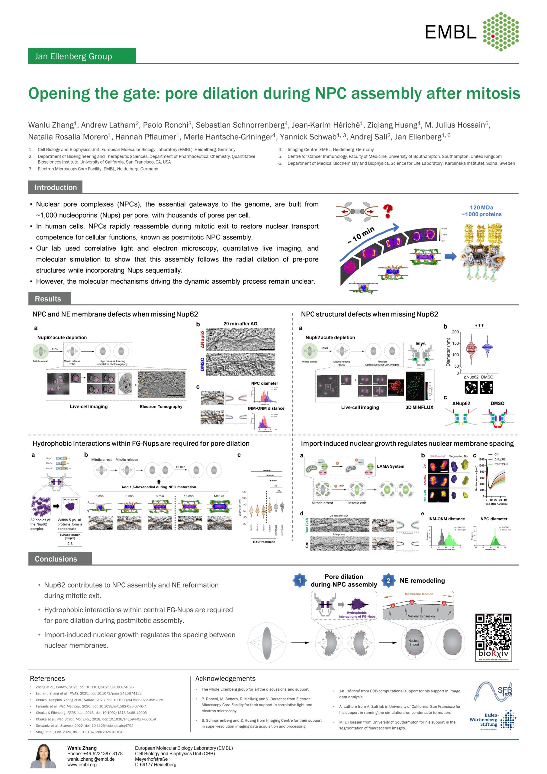
Opening the gate: pore dilation during NPC assembly after mitosis
Presenter: Wanlu Zhang
Authors: Wanlu Zhang, Andrew P. Latham, Paolo Ronchi, Sebastian Schnorrenberg,
Jean-Karim Hériché, Ziqiang Huang, M. Julius Hossain, Natalia Morero, Hannah
Pflaumer, Yannick Schwab, Andrej Sali, Jan Ellenberg

EMBL Heidelberg, Germany
Abstract:
Nuclear envelope (NE) reformation after mitosis is essential for daughter cell viability and requires tightly coordinated nuclear pore complex (NPC) assembly and nuclear membrane reformation. To reveal how these processes are mechanistically linked, we combined acute molecule perturbations in live cells with correlative 3D electron tomography or MINFLUX super-resolution microscopy. We show that degrading Nup62 during mitosis arrests NPC assembly at an intermediate step with smaller membrane pores and removes the whole central transport channel. Molecular dynamics simulations predicted that 32 copies of the central channel subcomplex, recruited into the previously unoccupied pore center, can self-associate via hydrophobic interactions to occupy the volume required for full pore size and exert an outward pushing force; indeed, disrupting these interactions during NPC assembly blocked pore dilation. Later in mitotic exit, perturbed cells exhibited impaired nuclear import, smaller nuclei, and looser NE spacing. Acute inhibition of nuclear import recapitulated these NE defects without affecting NPC assembly. Together, our findings reveal a new, two-step molecular mechanism linking NPC assembly and NE reformation. First, hydrophobic FG-nucleoporins dilate the assembling nuclear pore to its full width by forming the central transport channel, which then allows nuclear import-driven nuclear expansion leading to tight, regular NE membrane spacing.
The EMBO | EMBL Symposium ‘Seeing is believing: imaging the molecular processes of life‘ took place from 8 – 11 October 2025 at EMBL Heidelberg Advanced Training Centre and Virtual