Meet the best poster prize winners of ‘Seeing is believing: imaging the molecular processes of life’
At the beginning of October, the EMBO | EMBL Symposium ‘Seeing is believing: imaging the molecuclar processes of life’ took place and brought together the leading developers of imaging methods with cutting-edge applications that illustrate how imaging can answer biological questions.
Just before the conference there was an industry workshop organised by the industry sponsors of this event. The following four days featured six sessions, flash talks, poster sessions and lots of opportunities for networking.
We are pleased to present you the three poster prize winners of the event:
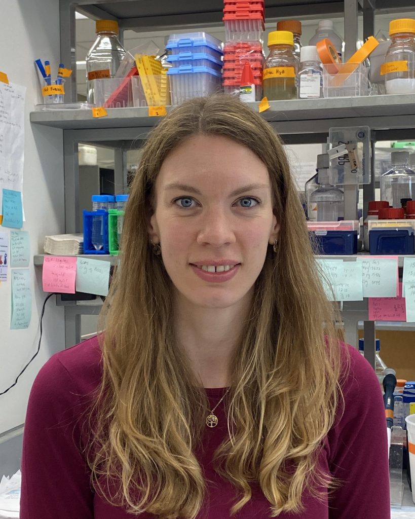
Visualizing endogenous Nanog behavior using a novel nanobody-based manipulation system
Presenter: Caroline Hoppe, Yale University School of Medicine, USA
Collaborators: Curtis Boswell, Alice Sherrard, Srikar Krishna Gopinath, Antonio Giraldez

In the developing zebrafish embryo, the pioneer transcription factor Nanog is required for genomic reprogramming and transcriptional activation. While recent work has suggested that Nanog coordinates the organization of large transcription bodies during the maternal-to-zygotic transition (MZT), the physiological relevance of these observations is unknown, as they are based on the live imaging of exogenous, fluorescently tagged Nanog. To probe the spatiotemporal dynamics of endogenous Nanog in vivo, we have developed a novel system termed GEARs: Genetically Encoded Affinity Reagents.
This system uses a collection of short epitope-based tags and their cognate (nanobody-) binders, which can
1) measure protein localisation and abundance and
2) perturb protein function in vivo.
We first validated the functionality of these reagents using exogenous reporters. After identifying the most suitable tag-binder pair, we generated endogenous GEAR-tagged nanog alleles with a rapid CRISPR/Cas9-based tagging pipeline. Using a GEAR-tagged Nanog, we then investigated its endogenous behaviour over the MZT. Strikingly, when visualizing Nanog localization and dynamics by live imaging, we find fewer Nanog clusters that are notably smaller in comparison to exogenously introduced Nanog further underscoring the physiological relevance of probing endogenous targets. To visualize these endogenous Nanog clusters at higher spatial resolution and to obtain further insights into the function of Nanog in controlling transcription and genome organization, we have also employed chromatin expansion microscopy (ChromExM) in conjunction with gene labelling. Together these results reveal principles of pioneer transcription factor function during genome activation and demonstrate the utility of the GEARS toolbox to provide a rapid and powerful strategy for interrogation of protein function while circumventing many challenges associated with conventional gene targeting.
Investigating contact formation and biophysical properties at the reconstituted immune synapse
Presenter: Franziska Ragaller, Karolinska Institutet and SciLife Lab, Sweden
Collaborators: Taras Sych, Luca Andronico, Adnane Achour, Erdinc Sezgin

The immune synapse is a spatiotemporally highly organized cell-cell contact between immune cells and their target cells, in which signalling events are tightly controlled. At this contact, the plasma membrane biophysical properties strongly influence the nature and dynamics of protein-protein interactions it harbours. This highlights the need for detailed investigation of biophysical properties alongside protein-protein interactions, to gain a comprehensive understanding of molecular mechanisms at the immune synapse.
Here, the immune synapse was reconstituted using model membrane systems alone or together with live cells. The use of synthetic membranes permitted investigation of defined lipid environments and specific ligand receptor pairs, which influence biophysical properties and contact formation at the immune synapse. The formation of the contact was investigated by confocal imaging and diffusion dynamics within the contact were examined by scanning fluorescence correlation spectroscopy (FCS). In summary, we could create a straightforward and widely applicable cell-cell contact reconstitution model, which allows the investigation of contact formation and biophysical properties such as diffusion dynamics of specific ligand-receptor pairs at the immune synapse.
Due to the confidentiality of the unpublished data, we cannot share the poster.
iNbSyt1: A highly versatile tool for perturbation-free live synaptic imaging
Presenter: Ronja Rehm, University Göttingen, Germany
Collaborators:
Karine Queiroz Zetune Villa Real, László Albert, Nikolaos Mougios, AmirMohammad Rahimi, Shama Sograte-Idrissi, Manuel Maidorn, Jannik Hentze, Markel Martínez-Carranza, Hassan Hosseini, Kim Ann Saal, Nazar Oleksiievets, Matthias Prigge, Roman Tsukanov, Pål Stenmark, Eugenio Fornasiero, Felipe Opazo

Synaptic vesicles (SVs) are essential organelles involved in neurotransmission, and their precise composition is necessary for information transfer from neuron to neuron. While many aspects of their functions at the synapse are known, open questions remain about how these organelles are generated and maintained to mature into functional SVs able of releasing neurotransmitters at the synapse.
In order to accurately study SV biogenesis, it is crucial to maintain the stoichiometry of SVs undisturbed. This renders experiments involving overexpression of a particular SV protein of difficult interpretation and calls for alternative techniques. A powerful approach to access the endogenous localization, distribution and trafficking of proteins are intrabodies: small probes based on high-affinity nanobodies that can be expressed inside cells. Here, I present the characterization of a family of affinity tools based on an intrabody against the SV Ca2+-sensor Synaptotagmin-1 (Syt1), named iNbSyt1. This intrabody allows not only the direct live imaging of SVs when fused to fluorescent proteins such as mScarlet and mNeonGreen, but also the detection of single action potentials when fused to a highly sensitive synaptically localized Ca2+-sensor, such as jGCaMP8s.
We have characterized this intrabody in detail, ensuring that its expression does not affect vesicle mobility, synaptic localization, or fusion capacity, making it a highly flexible tool to study synaptic processes in living neurons without genetic perturbation of the SV protein target. We also show that this affinity tool can be adapted to several super-resolution imaging modalities including STED microscopy, Structured Illumination Microscopy (SIM), DNA-PAINT and Expansion Microscopy.
Overall, a workflow to characterize and adapt a very specific nanobody to several different experimental needs is presented here to demonstrate how to best utilize these small binders and apply them in a wide range of imaging modalities
The EMBO | EMBL Symposium ‘Seeing is believing: imaging the molecular processes of life’ took place from 4 – 7 October 2023 at EMBL Heidelberg and virtually.