Edit

Tag:

brunner

Year
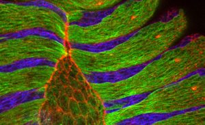
25 June 2009 The microscope image of the dorsal closure of a fly embryo shows alternating stripes of epithelial cells with aligned microtubule bundles (green) and epithelial cells treated with a microtubule-destroying drug (blue). Labelled in red is the protein actin that lines the border of cells, particularly the amnioserosa cells occupying the eye-shaped opening.

Uncovering how cells cover gaps

Science Researchers at the European Molecular Biology Laboratory (EMBL) in Heidelberg, Germany, came a step closer to understanding how cells close gaps not only during embryonic development but also during wound healing. Their study, published this week in the journal Cell, uncovers a fundamental…

2009

science

No matching posts found

EMBLetc.

Looking for past print editions of EMBLetc.? Browse our archive, going back 20 years.

EMBLetc. archive

Newsletter archive

Read past editions of our e-newsletter

For press

Contact the Press Office
Edit