Scientists, researchers, and publishers now have access to comprehensive checklists that provide unified guidelines for the publication of microscopy data and image analyses.
A working group of researchers from the Quality Assessment and Reproducibility for Instruments and Images in Light Microscopy (QUAREP-LiMi) initiative developed these checklists to improve the clarity, interpretability, and reproducibility of scientific image data and analyses in publications.
The paper aims to standardise and reinforce best practices for microscopy data sharing and see this approach perpetuated in future technical courses for life scientists and adopted by scientific journals within their own publication guidelines.
As part of a global initiative, researchers have drawn up guidelines for the publication of microscopy images in scientific outlets. The criteria, summarised in the form of checklists, form the basis for ensuring that published bioimaging data in the field of life sciences and medicine are intelligible and that the corresponding research is reproducible. This is the only way to unlock their full potential for research.
These results, achieved by 54 researchers from more than 48 institutes around the world and published in Nature Methods, are likely to influence global publication practices regarding microscopy images. The researchers make up a working group in a global initiative on Quality Assessment and Reproducibility for Instruments and Images in Light Microscopy (QUAREP-LiMi).
EMBL is globally known for its data and microscopy services and has long been a proponent of open access, which these guidelines help achieve by standardising how bioimaging data are shared.
Christian Tischer, a team leader in EMBL’s Data Science Centre, represented EMBL and played an integral role in formulating the checklists that are at the heart of the paper.
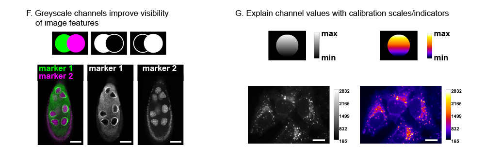
“To build upon published scientific results, it’s important that the data and corresponding analyses are scientifically accurate, reproducible, and accessible,” Tischer explained. “For microscopy-based research, this ranges from issues like the legibility of image data in publication figures, providing scale information, and a responsible choice of contrast adjustments, to sharing image data on public archives and making accessible the analysis pipeline on cloud computing platforms.”
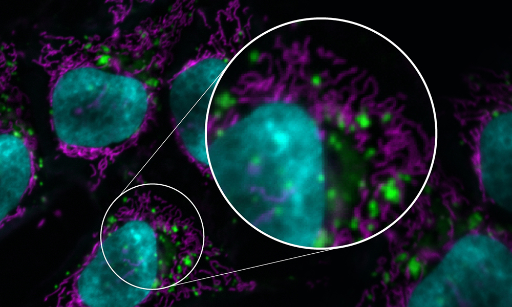
More than a million scientific papers are published in life sciences and medicine each year. Approximately one-third of them include images, such as microscopy data of cells or tissues. However, most of these images cannot be fully understood by the target audience, because key information is missing, e.g. information about scales used. Moreover, many lack information about how exactly the microscopy data were produced, preventing other researchers from reproducing comparable microscopy data.
Now, as part of the global QUAREP-LiMi initiative, this specialist working group developed communication guidelines, particularly for microscopy images and image analysis data.
“Scientists around the world and leading scientific journals have signalled an urgent need for publication standards for microscopy images,” said Helena Jambor, the initiator of the working group responsible for the guidelines, an author of the scientific paper, and a scientist at the National Center for Tumor Diseases Dresden (NCT/UCC) and at University Medicine Dresden. “These guidelines need to be drawn up by researchers because they know best which quality criteria are particularly important for their work. We have now succeeded in achieving a broad consensus, involving researchers from many of the world’s leading institutes in the life sciences.”
The group’s checklists offer some very specific guidance, for instance, making sure that relevant image sections are selected, that colour channels are named in fluorescence microscopy images, and that the colours chosen can be distinguished by readers who are colour-blind.
Knowing that many publications also present results of image analyses, the group prescribed that authors describe precisely how data are generated, for instance, which software solutions and settings were used, and that sample data are available to check the results. In general, images should be made available to the scientific community in suitable databases such that they can be used for further research.
“The guidelines are aimed at all researchers who work with light microscopy, from beginners to experts,” said Christopher Schmied, the research paper’s first author and a scientist at the Human Technopole Foundation in Milan and at the Leibniz-Forschungsinstitut für Molekulare Pharmakologie (FMP) in Berlin. “They enable the publication of images and image analysis results that meet high-quality standards, are reproducible and therefore plausible, and provide a good basis for further research projects.”
Within the checklists, the criteria are split into three levels so users can choose between minimal, recommended, and ideal requirements for good bioimaging data communication. “Our goal is for the criteria to be used by leading scientific journals as binding standards for publication,” Jambor said. “The chances of this are good. The members of the global initiative are constantly updating the checklists. And we will also be developing communication training materials and tutorials for microscopy images.”
“Our hope is that with continued adoption by scientists and scientific journals, we build a culture where life science research is able to progress more easily with the transparency that comes with sharing bioimaging data in a clear, consistent way,” Tischer said. “For people like me who also train life scientists on managing and analysing bioimaging data, we can propagate this information further in our courses as well.”
The QUAREP-LiMi initiative launched in April 2020 and now has nearly 550 members worldwide, including experts in bioimaging science and image analysis, microscope manufacturers and scientific publishers. The members are involved in 15 working groups that focus on specific topics. The working group responsible for drawing up the publication guidelines for microscopy images presented here is the Image Visualization and Analysis group, which is led by Jambor and Schmied and involves around 100 experts from all continents.
Modified from press release by Anna Kraft/NCT-UCC Dresden