A human fetal lung cell atlas uncovers proximal-distal gradients of differentiation and key regulators of epithelial fates
Cell 8 December 2022
10.1016/j.cell.2022.11.005
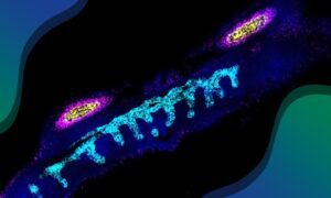
Researchers have created a spatial cell atlas of the developing human lung, describing 144 cell types/states and how they are regulated in unparalleled detail
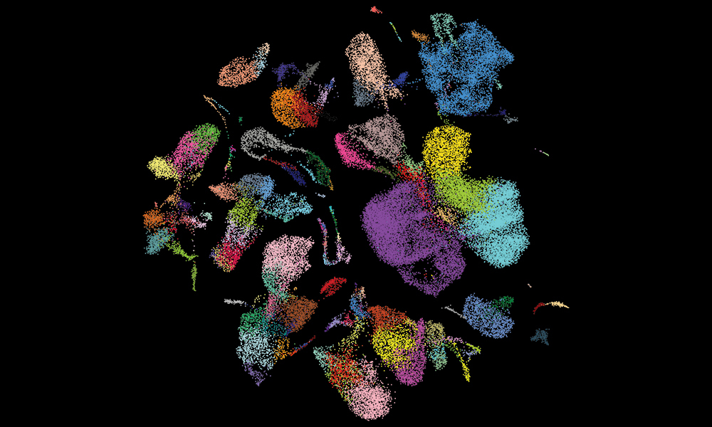
The developing human lung has been mapped in unprecedented detail, identifying 144 cell states in the early stages of life and uncovering new links between developmental cells and lung cancer.
In this latest study, which is part of the Human Cell Atlas Initiative to map every cell type in the human body, researchers at the Wellcome Sanger Institute, EMBL’s European Bioinformatics Institute (EMBL-EBI), the Gurdon Institute at the University of Cambridge, and collaborators, have examined which genes are activated in different stages of lung development, one cell at a time. They combined this with spatial technologies which pinpoint the exact location of cells to create the Developmental Lung Cell Atlas, showing how the respiratory system comes into being.
The atlas provides a unique and freely available resource that describes not only the individual cells that are created while the lungs are forming but also how these processes are controlled by genes.
This highly detailed atlas acts as a guidebook to healthy lung development and can be used as a baseline to investigate how lung diseases originate. It could also be used to create new models to study lung conditions and test potential therapies.
Many diseases have their origin during development. While single-cell mapping of adult organs in health and disease is unearthing new information about the lung, how the human lung forms in early life is still very much underexplored.
The early weeks of development are extremely dynamic, with cell types appearing and disappearing rapidly as the organ-building processes move between stages. To understand the development of the lung, it is necessary to have an atlas of all the lung cell types, including transient cells, at different stages of development.
To create this Developmental Lung Cell Atlas, researchers from the Wellcome Sanger Institute and collaborators combined single-cell sequencing of early-stage cells with spatial technologies to generate an in-depth dataset of lung development.
The team identified 144 cell types/states such as intermediate and transitional cell types, including a novel subtype that could be linked to the development of human small cell lung cancer later in life.
The team used the atlas to make predictions about how lung cells develop, especially which genes are the key players driving this process. They then used organoid models to test some of the key hypotheses, demonstrating that the atlas can be used to accurately predict the stages and cells involved in tissue development.
“Our high-resolution computational analysis has identified previously undescribed rare populations in the developing human lung, giving crucial insight into cell state transition that are not found, or not possible to investigate, in the adult organ,” said Peng He, Postdoctoral Fellow at EMBL-EBI and the Wellcome Sanger Institute.
In these new populations, we found one that is associated with a poorly understood type of small lung cell cancer in adults. While further research is required to understand the link, this shows the important insights into mechanisms shared by development and disease that can be found using our unique cell atlas. In addition to this, by including chromatin accessibility data and in vitro perturbation experiments, our work shows how key transcription factors can individually dictate lineage choices, allowing us to start to understand the regulatory logic that controls differentiation pathways.”
“A complete Human Cell Atlas, detailing all stages of life, will help explain many aspects of human health and disease,” said Sarah Teichmann, Head of Cellular Genetics and Senior Group Leader at the Wellcome Sanger Institute, and Co-Founder of the Human Cell Atlas. “This includes the cells that are the building blocks of organs as they form in early life, which we lose as an adult. Our Developmental Lung Cell Atlas allows us to look at the stages and pathways that adult lung cells have gone through, and how these beginning phases influence disease later on. This is an important contribution towards the first draft of the Human Cell Atlas.”
Read the full press release on the Sanger Institute website.
This study is part of the international Human Cell Atlas (HCA) consortium, which is aiming to map every cell type in the human body as a basis for both understanding human health and for diagnosing, monitoring, and treating disease. An open, scientist-led consortium, HCA is a collaborative effort of researchers, institutes, and funders worldwide, with more than 2,500 members from 85 countries across the globe.
These data sets are available for interactive analysis.
Developmental tissue was provided by the Human Developmental Biology Resource (HDBR), which provides human embryonic and fetal tissues to ethically approved scientific studies such as the Human Developmental Cell Atlas to enable research into understanding human development to help improve health.
This research was part-funded by Wellcome, NIHR Cambridge Biomedical Research Centre, the MRC, EMBL and others. A full acknowledgements list is available in the paper.
Cell 8 December 2022
10.1016/j.cell.2022.11.005