Cells eat themselves into shape
Specialised endocytocis consumes membrane tendrils
In a nutshell:
– Endocytosis can play key role in changing cells’ shape
– To quickly smooth out their surface, cells in fruit fly embryo ‘suck in’ long tubes of membrane
– Discovered thanks to new imaging strategy and combination of light and electron microscopy
The process cells use to ‘swallow’ up nutrients, hormones and other signals from their environment – called endocytosis – can play a crucial role in shaping the cells themselves, scientists at the European Molecular Biology Laboratory (EMBL) in Heidelberg, Germany, have found. The study, published today in Nature Communications, could help explain how the cells on your skin become different from those that line your stomach or intestine.
“We’re the first to show that endocytosis really drives changes in cell shape by directly remodelling the cell membrane,” says Stefano De Renzis, who led the work.
De Renzis and colleagues made the discovery by studying the fruit fly Drosophila,which starts life as a sac. The fly’s embryo is initially a single large cell, inside which nuclei divide and divide, until, at three hours old, the cell membrane moves in to surround each nucleus, so that in about an hour the embryo goes from one to 6000 cells. As this happens, cells change shape. The cell membrane starts off with lots of finger-like tendrils sticking out of the embryo, and in about 10 minutes it smoothes down to a flat surface, like a rubber glove transforming into a round balloon.
Video 1: How one becomes 6000: cellularisation in Drosophila embryo
The EMBL scientists found that, for this quick shape-shift to happen, the cells ‘eat up’ their finger-like offshoots. And, to quickly take up all that excess membrane, cells adapt their ‘feeding strategy’. Instead of bending a little pouch of membrane inwards and eventually detaching it into the cell as a round pod, or vesicle, the fruit fly embryo’s cells suck in long tubes of membrane. Once inside the cell, those tubes are then processed into smaller vesicles.
Video 2: Cells eat themselves into shape
The findings, which include uncovering one of the key molecules involved, provide a new way of thinking about how cells take on the shape required to perform different tasks – and not only in fruit flies.
“This outward-facing – or apical – surface is the main difference between different kinds of epithelial cell,” says De Renzis. “The cells on your skin are smooth, but the ones lining your intestine have lots of ‘fingers’ like our fly embryos, and we know for instance that some bowel diseases involve problems in those ‘fingers’.”
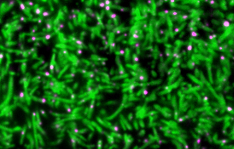
For this work, Piotr Fabrowski in De Renzis’ lab developed a new strategy for imaging the fruit fly embryo and Aleksandar Necakov, a joint post-doctoral fellow in the De Renzis and John Briggs labs at EMBL, combined light and electron microscopy to see how different the swallowed membrane tubes are from the vesicles usually formed in endocytosis.
Further information
Combining light and electron microscopy, by Briggs and Kaksonen labs
EMBL Interdisciplinary Post-docs (EIPOD) initiative


