
Muscular worm larva
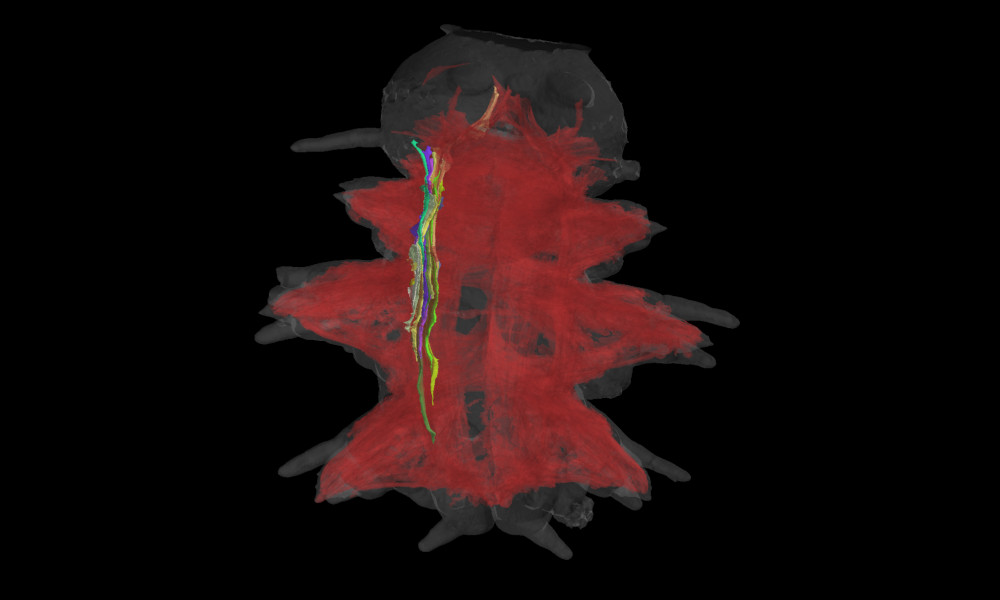
The image shows a larva of Platynereis dumerilii, a marine worm, with all its muscles highlighted. These worms populate coastal marine waters from temperate to tropical zones. Platynereis is an important lab animal and is used in many phylogenetic studies as a model organism.
The image here was produced by Constantin Pape, a visiting predoctoral fellow in the Kreshuk group at EMBL Heidelberg. He used data from an electron microscope and rendered the muscles via an automated analysis. Using this image he tried to learn more about the correlation between morphology and gene expression in Platynereis dumerilii.
The grey outline shows the larva’s body, while red highlights the muscle strands. The muscles of one individual strand are highlighted in different, brighter colours.
Credit: Constantin Pape/EMBL
If you have a stunning picture of your science, your lab or your site, you can submit it here.


