Colloidal Coomassie Staining
Protocol
Empowering research through mass spectrometry-based proteomics
We only perform processing of gel bands stained with Coomassie. Therefore, please ensure your Coomassie stain is MS-compatible by consulting the manufacturer’s datasheet or contacting their customer service. If you want to mix your own staining solution, please see protocol on the right for a compatible recipe.
Important information:
Protocol
For external customers:
When submitting gels from outside EMBL Heidelberg, we prefer that you send already excised gel bands. This will minimize the risk that your sample(s) will get damaged during transport.
Please refer to the protocol on the right to excise the gel bands.
For customers from EMBL Heidelberg:
You can bring your gel to our facility after destaining.

Protocol
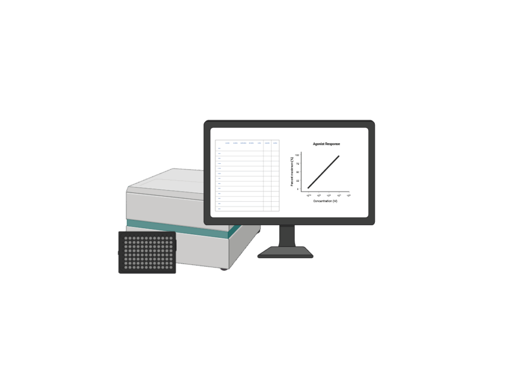
Based on the method described by Wiśniewski and Gaugaz (DOI: 10.1021/ac504689z), this fluorescence-based assay is cost-effective, user-friendly, and compatible with a wide range of sample types and buffer components.
Note: This assay requires black microplates and a fluorescence-capable plate reader. Please check compatibility with your equipment.
Protocol
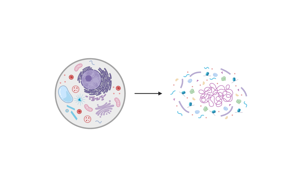
This protocol describes a simple and efficient denaturing lysis method for mammalian cells. It is intended as a general option when no project-specific protocol is available.
If you are uncertain whether this procedure is suitable for your experimental setup or research question, please contact us for guidance.
Protocol