
James Swoger
Head of Mesoscopic Imaging Facility
ORCID: 0000-0003-3805-0073
Edit
Head of Mesoscopic Imaging Facility
ORCID: 0000-0003-3805-0073
EditThe Mesoscopic Imaging Facility at EMBL Barcelona was established to advance imaging techniques specialized in studying tissue biology. Located within the Barcelona Biomedical Research Park (PRBB), alongside EMBL Barcelona’s independent research groups, our facility provides cutting-edge mesoscopic imaging technologies tailored for research in developmental biology and disease modeling.
We are an interdisciplinary, international team with expertise in biology, engineering, and physics, dedicated to offering state-of-the-art imaging capabilities to scientists worldwide.
Our facility plays a key role in supporting fundamental research by enabling high-resolution, large-scale imaging of complex biological systems. We provide advanced microscopy solutions for studying dynamic processes in living tissues, such as embryonic development, organogenesis, and cellular interactions in three-dimensional environments. With expertise in imaging large and delicate specimens, we assist researchers in designing optimal imaging workflows, from sample preparation to data acquisition and analysis, ensuring that they can capture biological phenomena with unprecedented clarity and precision.
Beyond fundamental research, we are actively exploring new applications that extend the impact of mesoscopic imaging into clinical and translational fields. Our facility is at the forefront of 3D histopathology, developing light-sheet microscopy approaches that enable high-resolution, volumetric imaging of tissue samples without sectioning—offering a powerful alternative to conventional histological methods. Additionally, we are pioneering live organoid imaging, providing researchers with dynamic insights into cellular behavior, tissue organization, and disease progression in physiologically relevant models. By bridging the gap between advanced microscopy and biomedical applications, we aim to empower hospitals, clinicians, and researchers with novel imaging solutions that can revolutionize fundamental, diagnostics and therapeutic research.
Our facility is strategically positioned near the Advanced Light Microscopy Unit (ALMU) at the Centre for Genomic Regulation (CRG), enabling seamless access to complementary imaging modalities. We also foster strong collaborations with leading research institutes on campus and across the region, enhancing our ability to support groundbreaking biomedical discoveries.
Find out more about our facility in an interview by El·lipse.
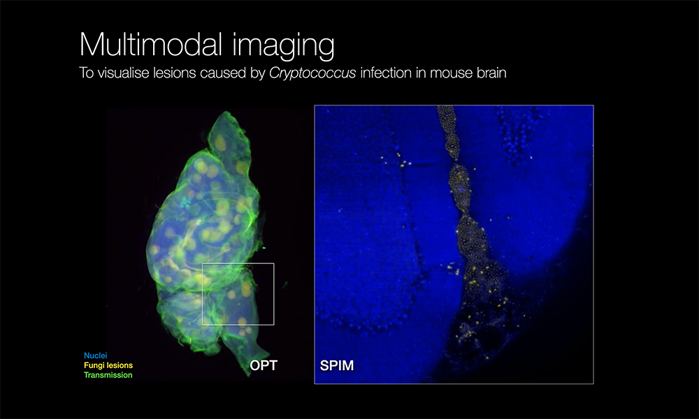
We collaborated with the lab of Greetje Vande Velde from KU Leuven to image cryptococcal lesions in intact brain tissue using cleared sample light-sheet microscopy. This 3D imaging approach revealed the spatial organization of infections, advancing our understanding of fungal pathogenesis.
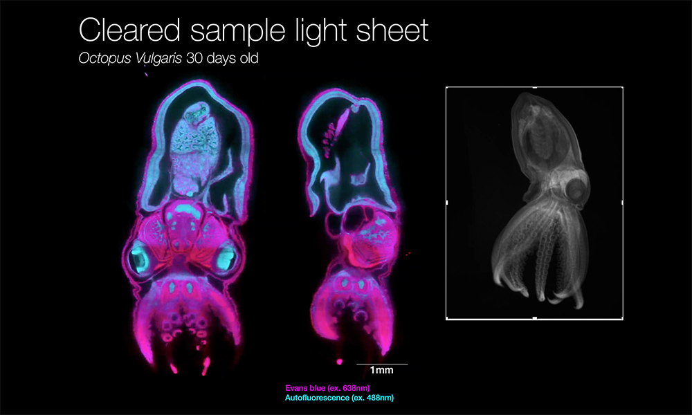
We collaborated with ICM-CSIC to study the digestive system development of Octopus vulgaris paralarvae using tissue clearing and light-sheet fluorescence microscopy. This 3D imaging approach provided insights into ontogenetic and evolutionary trends in cephalopod digestive systems.
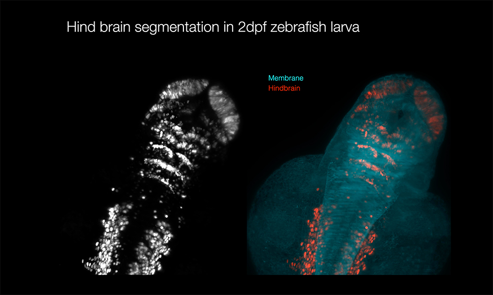
We collaborated with UPF researchers to investigate the neurogenic fate of hindbrain boundaries during development. Using light-sheet microscopy, we visualized the 3D organization of these regions, providing insights into their role in neurogenesis.
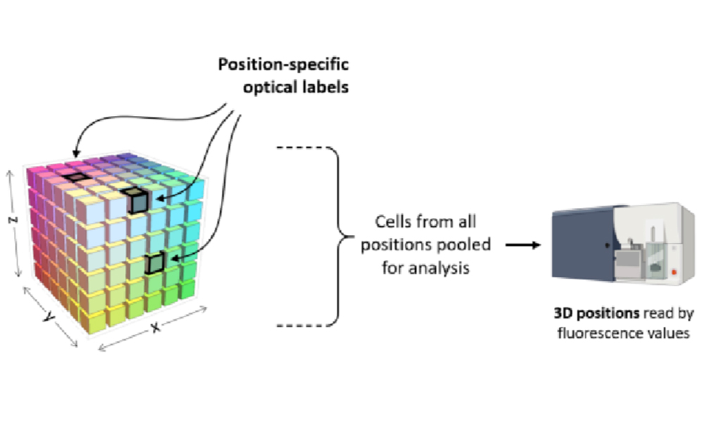
With the Sharpe Lab, we developed C3PO, a novel method that optically encodes 3D cell positions prior to tissue dissociation. This technique enables the reconstruction of spatial transcriptomic maps from single-cell data, bridging the gap between high-resolution imaging and omics.