James Swoger
Head of Mesoscopic Imaging Facility
ORCID: 0000-0003-3805-0073
Edit
Head of Mesoscopic Imaging Facility
ORCID: 0000-0003-3805-0073
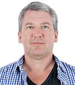
EditJim Swoger completed his PhD at McMaster University in Hamilton, Canada, in 1997, with a thesis on semiconductor laser physics. He then moved to the Light Microscopy Group at EMBL in Heidelberg, Germany, where he worked on optical microscopy systems such as confocal theta microscopy, multi-imaging-axis microscopy and selective plane illumination microscopy.
In 2005 he joined the Sharpe lab at the Human Genetics Unit of the Medical Research Centre in Edinburgh, UK, and spent a year investigating 3D imaging of mouse embryology using optical projection tomography.
From 2006 to 2017 he was a staff scientist at the Centre for Genomic Regulation in Barcelona, Spain, where he developed optical techniques for mesoscopic imaging in the Systems Analysis of Development group. He has been the Head of the Mesoscopic Imaging Facility at EMBL Barcelona since October 2017.

Project Manager
ORCID: 0000-0002-0154-9455
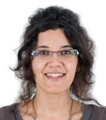
EditGopi completed her PhD at the Max Planck Institute for Cell Biology and Genetics (MPI-CBG) in Dresden, Germany, in 2016, where she worked with an interdisciplinary team to develop light sheet microscopes capable of long-term, high-throughput imaging of fragile samples and real-time image-processing. Using these systems, she focused on studying germlayer dynamics during zebrafish development.
Prior to that, at the Tata Institute of Fundamental Research (TIFR), Mumbai, India, Gopi used confocal microscopy for live cell imaging, FRAP and sub-cellular laser ablation assays to understand cell dynamics during Drosophila embryogenesis.
In 2016, Gopi joined the light microscopy core facility at the Cancer Research UK Cambridge Institute (CRUK CI) in Cambridge, UK, as a Senior Scientific Associate, where she established light sheet microscopy as a tool for imaging human and mouse tumour organoid models in 4D. She also participated in designing the institute’s image data-handling infrastructure and taught courses on open-source image processing tools.
With expertise in a wide range of imaging and biological systems, Gopi joined the Mesoscopic Imaging Facility at EMBL Barcelona in October 2018 as a Project Manager.

Mesoscopic Imaging Specialist
ORCID: 0000-0003-0169-4107
EditMontse has always had a genuine interest in bringing to light small details of life through images. After completion of her degree in biology at the Universitat Pompeu Fabra (UPF), she spent two years doing research on inner ear neurogenesis in the Developmental Biology Research Group (UPF).
She then moved to marine microbiology, and in 2013 she completed a PhD, with a thesis titled “Gene Expression in Marine Microorganisms”, in which she used genomic and molecular approaches to understand the ecology of marine microbial plankton (Universitat Politècnica de Catalunya/Institute of Marine Sciences, ICM-CSIC). During her PhD she had the opportunity to participate in the Tara Oceans Expedition on board the Tara schooner, studying the microbial diversity of the marine ecosystem. After her PhD, she was involved in photography, time-lapse photography, and video projects.
In 2017, Montse joined the Sharpe lab in the Centre for Genomic Regulation in Barcelona and gained experience in a wide range of clearing methods for rendering tissues transparent. Montse joined the Mesoscopic Imaging Facility at EMBL Barcelona in October 2017.
Imaging Technology Specialist
EditAfter his BEng studies in Electrical and Electronic Engineering at the University of Strathclyde, Spiros Bakas completed his MSc degree, in 2016, at the University of Glasgow in the subject of Nanoscience and Nanotechnology.
In 2017, he returned to the University of Strathclyde to start his PhD with a thesis in the miniaturization of light sheet microscopy using MEMS for all-optical control. During his PhD he had the opportunity to visit the IIT Gandhinagar, India, where he collaborated with research students on the development of a miniaturized light sheet system based on his PhD work. His research has focused on finding the methods to setup an imaging system in a compact footprint, with the use of original 3D printed opto-mechanics, while developing the synchronization protocol of the active elements that allow 3D imaging without a translation or rotational stage.
Since January 2023 he has joined the Mesoscopic Imaging Facility at EMBL Barcelona as an Imaging Technology Specialist to apply his expertise on the development of novel microscopy systems.

Image Analysis Specialist
ORCID: 0000-0002-7637-4258
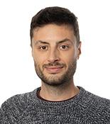
EditAfter his studies in theoretical physics at the University of Milan-Bicocca (Italy), Nicola joined the Atomic and Molecular Physics Institute (AMOLF) in Amsterdam, the Netherlands, where he completed his PhD in 2017 in the lab of Dr. Jeroen van Zon. Nicola developed a time lapse microscopy technique to perform long-term study of the roundworm C. elegans, and used an interdisciplinary approach combining microfabrication techniques, microscopy development and image analysis.
After obtaining a Human Frontier Science Program fellowship, Nicola started his postdoc in 2018 in the lab of Prof. Jan Huisken at the Morgridge Institute for Research in Madison (WI, USA), where he developed a method to improve optical penetration and image quality of Zebrafish and Drosophila embryos using a combination of visible and near infrared light sheet fluorescence microscopy and deep learning algorithms.
In 2019, Nicola joined the group of Dr. Vikas Trivedi at EMBL Barcelona as postdoc fellow. There, he pursued his interests in the early development of multicellular organisms using embryonic organoids from zebrafish and mouse stem cells. Nicola developed a semi-automated image analysis software (MOrgAna) that allows to perform morphometric and fluorescence quantification of hundreds of organoid images using machine learning.
Equipped with the image analysis expertise acquired during his scientific career, Nicola joined the Mesoscopic Imaging Facility at EMBL Barcelona in November 2021 as Image Analysis Specialist.