Previous and current research
Cells organise their biochemical reactions into functionally distinct compartments. An increasing number of studies report that many cellular compartments are not membrane enclosed, but rather assemble dynamically by phase separation. membrane-less compartments occupy an intermediate length scale between the nanoscale of individual macromolecules and the microscale of cells. They constitute non-deterministic and non-stoichiometric assemblies. A major theme of our research is to harness cutting-edge in-cell structural biology to unravel functional molecular architectures underlying cellular compartmentalisation by phase separation.
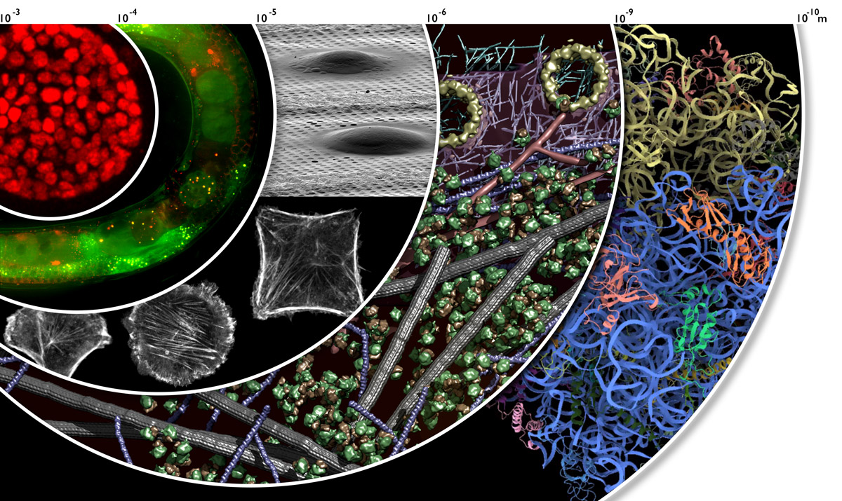
Cryo-electron tomography (cryo-ET) of intact cells, in combination with correlative approaches, is uniquely positioned to provide structural information across scales, from whole organisms to individual macromolecules. Our recent work on sample preparation shows that we can now prepare site-specific ‘electron-transparent windows’ in suitable eukaryotic systems, which allow direct examination of the structural features of cellular compartments and macromolecular assemblies in their native, functional environment. Cryo-focused ion beam (cryo-FIB) micromachining literally opened these electron-transparent windows into cells, which are otherwise too thick to be examined at high resolution in a transmission electron microscope. Now, cryo-FIB micromachining is reliably applied to a wide variety of single-cell cultures. With the understanding that processes inherent to metazoans (i.e. developmental and tissue-specific biology) often can’t be recapitulated in 2D cell culture models, we developed a cryo-lift-out strategy for FIB preparations of voluminous specimens. To attain precise cryo-FIB targeting of rare and dynamic assemblies, we developed 3D cryo-correlative light and electron microscopy. We harness this technology to elucidate molecular architectures underlying stress granules in HeLa cells, which are RNA bodies that form rapidly in the cytoplasm upon cellular stress; and centrosomes, which are sites of microtubule nucleation, in developing C. elegans embryos. We combine these studies with a quantitative description of the crowded nature of the cytoplasm and of its local variations, to provide a direct readout of the impact of excluded volume on molecular assembly in living cells.
Future projects and goals
Our team continues to advance techniques for in-cell structural biology. New lines of research build on our first successful steps towards the transformative possibility of imaging macromolecular complexes inside cells at near-atomic resolution, and directly reveal how the fine compositional and conformational states of these complexes are linked to biological functions. With this enabling technology, we tackle questions spanning a broad spectrum of biological complexity, from the simplest bacterium as a blueprint for the most conserved molecular machinery, to translationally relevant molecular architectures in human induced pluripotent stem cells and 3D organoids. Supported by our outstanding research environment at EMBL, we increasingly integrate omics and systems biology approaches into our research, to transform our structural discoveries into mechanistic understanding.