Get to know the best poster prize winners of ‘In situ structural biology: from cryo-EM to multi-scale modelling’
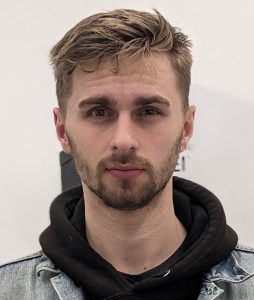
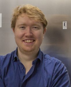
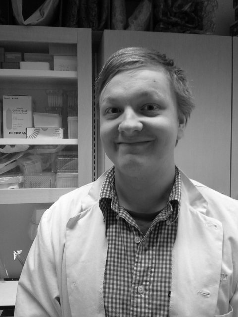
161 on-site and 66 virtual participants from all around the world attended the EMBO Workshop ‘In situ structural biology: from cryo-EM to multi-scale modelling’ which was held on 8 – 11 February 2023 in Heidelberg. From 68 posters that were on display around the ATC’s helices, participants could vote for their favorite posters. We are glad to announce now the five poster prize winners: congratulations to Joe, Matthew, Max, Sven, and Veijo!
Towards visual proteomics of gene expression in a minimal bacterium
Presenter: Joe Dobbs, EMBL Heidelberg, Germany

Recent advances in cryogenic electron tomography (cryoET) are now allowing researchers to structurally characterize protein complexes in situ at increasingly higher resolution, investigate interaction networks and protein communities, and derive functional insight from assemblies unsuitable for traditional structural methods. We are investigating the molecular sociology of gene expression, focusing on RNA polymerase in the minimal prokaryote Mycoplasma pneumoniae. M. pneumoniae has been used recently by our group in studies of the ribosome, providing the first detailed snapshots of translation structural dynamics inside the cell. RNA polymerase represents an intermediate sized (400 KDa), abundant (~300 copies/cell), molecular interaction mapped complex we aim to take to high resolution by way of improving particle identification and by using new tools and workflows. This census of RNA polymerase in M. pneumoniae can then be combined with the new structural understanding of translation in this minimal bacterium. However, as we target increasingly small objects of interest, cryoET’s existing challenges are critical impediments. To address these we are developing a correlative cryogenic single molecule localization microscopy (SMLM) approach to precisely identify specific proteins and complexes, and will push the limits of complex size for in situ cryoET and subsequent structure determination to investigate cellular communities of molecular machines at high resolution.
Due to the confidentiality of the unpublished data, we cannot share the poster.
Visualizing the ultrastructure of the dopaminergic synapse using cryo-CLEM and cryo-ET
Presenter: Matthew Lycas, EPFL, Switzerland

The dopaminergic synapse is critical for controlling the release of dopamine, a neurotransmitter responsible for motivation and movement among many other biological roles. Yet, the knowledge of the architecture and organization of the intact dopaminergic synapse has been limited. We have developed an optimized method for growing dopaminergic neuronal cultures on cryo‑EM grids allowing for imaging of synapses in primary cultures with Cryo‑CLEM. Using an AAV expression system, we expressed mCherry only in dopaminergic neurons allowing us to identify the dopaminergic neurons in the mixed culture through spinning disk confocal microscopy of the cryo‑preserved samples. Using Cryo‑CLEM, we obtain nanometer resolution atlases allowing for qualitative and quantitative description of dopaminergic axons and synapses. We were also able to observe and quantify changes in the synaptic physiology by subjecting the cultures to dopaminergic pharmacological manipulation prior to cryo‑fixation. Finally, tomography was performed at dopaminergic synapses, allowing for detailed segmentations and in situ protein structure determination. Together, these methods demonstrate the full pipeline for characterizing ultrastructural changes of dopaminergic synapses at subnanometer resolution.
Methodological advances in cross‑linking mass spectrometry increase detection sensitivity and quantification accuracy for large‑scale interactomics studie
Presenter: Max Ruwolt, FMP, Germany

Cross‑linking mass spectrometry (XL‑MS) is a powerful method for the investigation of protein‑protein interactions (PPIs) from highly complex samples. Improving the sensitivity of cross‑link detection and advancing the accuracy of cross‑link quantitation have been two essential aspects in the XL‑MS field. Over the past years, we have been developing advanced methods aiming both important aspects. First, we developed a real‑time library search assisted workflow to improve the sensitivity of cross‑link identification. We use a set of diagnostic peaks to distinguish cross‑links from mono‑links and apply real‑time library search to target only potential cross‑links for MS2 sequencing. Using this acquisition strategy, we increase the number of cross‑linked spectrum matches by 35% while reducing mono‑linked spectrum matches by 88% compared to standard acquisition with a 1 h gradient. Second, we develop tandem mass tag (TMT)‑based XL‑MS workflow to enable efficient and robust cross‑link quantification. We construct a two‑interactome dataset by spiking‑in TMT‑labeled cross‑linked E. coli lysate into TMT‑labeled cross‑linked HEK293T lysate to assess the efficacy of cross‑link identification and accuracy of crosslink quantification using different MS acquisition strategies. We show that a MS2 method with stepped collision energies 42 ± 6 provides a vast number of quantifiable cross‑links with high quantification accuracy. Taken both efforts together, we made significant contributions in the method development of XL‑MS. We are able to increase detection sensitivity and quantification accuracy of cross‑links which allow us to provide additional information on protein conformations, dynamics and PPIs.
Revisiting visual proteomics by cryo electron tomography of cryo FIB lamellae
Presenter: Sven Klumpe, Max Planck Institute of Biochemistry, Germany

Cryo electron tomography of cryo focused ion beam(FIB)prepared lamellae has enabled unprecedented insights into cellular ultrastructure and paved the way for in situ structural biology of large macromolecular complexes. Limitations remain in the form of relatively low throughput, limited and slow targeting capabilities, and the need for user input in many steps along the way, e.g., lamella site selection. Although both academic and commercial software for automation are now available, the throughput of lamella preparation required for “visual proteomics” of low copy number proteins and complexes is still not met. It is limited by all the steps that need to be performed by the microscope operator in e.g. choosing lamella sites or correlating fluorescencelight microscopydata. Using novel cryo FIB instrumentation, namely an automated sample exchange,in chamber light microscopes, and plasma ion sources,and exploiting machine learning models, we demonstrate a fully automated workflow for lamella preparation from sample loading to unloading. This enables the preparation of 12 cryo FIB prepared grids in a single session. We show that as little as 40 images are sufficient to train U Net based models capable of selecting suitable lamella positions and enabling the growth of datasets during sample preparation in anhuman in the middle approach. Furthermore, asthere is evidence that the models are transferable (e.g. a model trained on C. reinhardtii data is capable to produce lamellae from S. cerevisiae samples), the method holds the potential for the intriguing idea of an universal model for cellular lamella preparation. Automated selection and preparation of lamellae as well as automated sample exchange is implemented in the open source software package SerialFIB. In combination, these improvementsmake sample preparation of cells for cryo electron tomography without user input possible. Finally, we discuss preliminary data on how novel workflows and similar computer vision approaches can aid in fluorescence targeting and lamella throughput from high pressure frozen tissue such as D. melanogaster egg chambers and C. elegans larvae by on grid milling or cryo lift out.
Elucidating the nanoscale architecture of ER LD contact sites via cryo correlative light and electron tomography
Presenter: Veijo Salo, EMBL Heidelberg, Germany

Lipid droplets (LDs) are neutral lipid (NL) storage organelles vital for metabolism. LD biogenesis occurs in the endoplasmic reticulum (ER) in a process orchestrated by a molecular machinery that includes the lipodystrophy protein seipin, its interactor LDAF1 and the LD coat protein perilipin 3. Here, we use cryo correlative light and electron microscopy to construct 3D structural models representing the timeline of LD biogenesis in human cells. Cells with key LD proteins tagged endogenously with fluorescent markers are stimulated towards LD formation and frozen in near native state. They are thinned by focused ion beam milling, imaged by cryogenic Airyscan microscopy to localize LD biogenesis sites and targeted for in situ cryo electron tomography. For nanoscale precision localization of seipin mediated ER LD contacts and LD formation sites in the cryo tomograms, we further developed genetically encoded nanoparticles that tether to seipin upon drug induction.
Our results reveal key features of seipin mediated ER LD membrane continuities. The diameter of these high curvature necks is restricted in a seipin dependent manner to ~20 nm, with the outer leaflet of the ER clearly continuous with the LD monolayer. This neck architecture suggest seipin adopts a more open conformation compared to in vitro structures. In ~50% of ER LD contacts, the ER lumenal leaflet protrudes into the LD, forming a bubble like structure. We hypothesize that the bubbles serve as a mechanism to control LD growth and entail a further conformational change of the seipin oligomer. Going forward, our pipeline will provide direct observations to allow construction of comprehensive 4D models of LD biogenesis.
Due to the confidentiality of the unpublished data, we cannot share the poster.