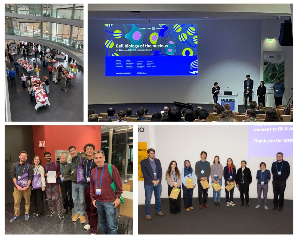
Meet the poster prize winners of ‘Cell biology of the nucleus’
The inaugural edition of the EMBO | EMBL Symposium ‘Cell biology of the nucleus’ took place in Heidelberg from 18 – 21 November 2025 and brought together scientists from a range of backgrounds working on different aspects of the nucleus, including nuclear architecture, nuclear envelope remodeling, nuclear transport, nuclear mechanics, and more.
The meeting welcomed 165 participants on site and 59 online, with 9 fellowships provided by the EMBL Corporate Partnership Programme and EMBO. The participants connected and discussed topics such as the evolution of the nucleus, biophysics and mechanics of the nucleus, dynamic nuclear architecture, nuclear signaling and communication, cell divison and the nucleus, and more while the key focus of this conference was to showcase the cell biological mechanisms that define the nucleus as an organelle.
Several networking and career planning opportunites were offered during ‘meet the speakers’ or ‘meet the editiors’ sessions, and participants could present their posters during two poster sessions.
In total, 93 posters were displayed; offering a broad snapshot of current research. From these, five poster prize winners were selected, and we are pleased to introduce four of them and their work in this post.
The AAA+ ATPase Torsin sustains CLCC1 function to promote NPC biogenesis
Presenter: Daria Maslennikova
Authors: Daria Maslennikova, Harry Baird, Jose Maria Mateos Melero, Alessia Loffreda, Caroline Ashiono, Federico Uliana, Madhav Jagannathan, Ulrike Kutay
ETH Zürich, Switzerland
Abstract:
Primary dystonia is a severe neurodevelopmental disorder characterized by involuntary muscle contractions, most often caused by a point mutation in Torsin1A. Torsins are the only
AAA+ ATPases localized to the lumen of the endoplasmic reticulum (ER) and the contiguous nuclear envelope (NE). They have been implicated in lipid metabolism, nuclear pore complex (NPC) biogenesis, and protein secretion, yet their molecular function and substrates have remained enigmatic. We have used male dtorsin D. melanogaster knockout flies, which are sterile, as a model to study Torsin function in germline development. We found that dtorsinKO flies exhibit pre-meiotic germ cell arrest and display NE herniations containing stalled NPC assembly intermediates at their constricted necks. Interestingly, NPCs accumulated in cytoplasmic membrane stacks resembling annulate lamellae (AL), indicating that dTorsin is specifically required for NPC biogenesis at the NE. To gain insight into Torsins’ molecular function in NPC biogenesis, we used proximity labeling in human cells, identifying the ER transmembrane protein CLCC1 as a prominent human Torsin1A interactor. We show that CLCC1 depletion phenocopies Torsin loss-of-function, both in human cells and fly germline cells lacking the Drosophila orthologue of CLCC1. While wild-type dTorsin rescues dtorsinKO defects in the Drosophila germline, mutating key residues at the predicted dTorsin–dClcc1 interface failed to rescue NPC biogenesis. Remarkably, overexpression of Drosophila Clcc1 (dClcc1) rescued NPC biogenesis, germline development and fertility in dtorsinKO flies, indicating that dTorsin is mainly required to sustain CLCC1 functionality. Immunogold EM revealed enrichment of CLCC1 and Torsin1A at the apical regions of NE herniations, suggesting their role in fusion of the inner (INM) and outer nuclear membranes (ONM) during NPC maturation and guiding models of how Torsin and CLCC1 act together during INM–ONM fusion. Our work identifies CLCC1 as a key Torsin interactor and a potential therapeutic target in early-onset dystonia.
Due to the confidentiality of the unpublished data, we cannot share the poster.
Characterization of the nuclear pore complex in Entamoeba histolytica
Presenter: Huda Amilina
Authors: Huda Amilina, Herbert J Santos, Tomoyoshi Nozaki

The University of Tokyo, Japan
Abstract:
The emergence of the nucleus marked a major transition in eukaryotic evolution, creating a
physical barrier that separates genetic material from the cytoplasm. This compartmentalization required transport systems to mediate communication between the nucleus and cytoplasm. Central to this exchange are nuclear pore complexes (NPCs), the sole bidirectional gateways for proteins, RNAs, and other macromolecules. NPCs are large assemblies of more than 30 nucleoporins (Nups), organized into subcomplexes that are broadly conserved across the six major eukaryotic supergroups. Despite this conservation, structural and compositional variations highlight lineage-specific innovations, particularly in protists that diverged early in eukaryotic history.
Here, we investigate the NPC of Entamoeba histolytica, an Amoebozoan whose evolutionary position provides insight into both conserved and divergent NPC features. This parasite is also the causative agent of amebiasis, a neglected tropical disease responsible for an estimated 80,000 deaths annually, underscoring both its biomedical and evolutionary significance. Using the highly conserved FG-repeat nucleoporin EhNup98 as bait in pull-down assays, we identified interacting Nups by mass spectrometry, including a previously uncharacterized protein resembling Nup53/Nup35 in Opisthokonts. Comparative analysis revealed that this Nup53-like protein contains a predicted RNA recognition motif (RRM) domain that, although poorly aligned at the sequence level, is structurally conserved across lineages. In addition, it harbors a repeat-rich region absent in other eukaryotes and a truncated C-terminal domain, typically required for binding Nup155, suggesting altered interaction networks within the E. histolytica NPC. Gene knockdown further demonstrated that both EhNup98 and EhNup53 are essential for parasite viability, highlighting their central roles in NPC integrity. Together, these findings delineate a conserved NPC core while uncovering lineage-specific innovations, providing an important step toward understanding the nuclear architecture of E. histolytica and offering insights into both cell biology and the evolutionary history of the nucleus.
Due to the confidentiality of the unpublished data, we cannot share the poster.
Poster prize kindly sponsored by FEBS Letters
Histone deacetylation drives apoptotic chromatin compaction to limit shedding of DNA from the dying cell
Presenter: Maximilian F.D. Spicer
Authors: Maximilian F. D. Spicer, Sanne Wijma, Nikki Schuette, Daniel Gerlich
Institute of Molecular Biotechnology, Austria
Abstract:
Chromatin compaction regulates genome accessibility, shaping gene expression and cellular identity throughout the life of a cell. During apoptosis, genome-wide compaction converts a mixture of open and closed interphase chromatin into one coalesced, dense mass. This is thought to suppress immune stimulation by DNA outside the nucleus, however both the mechanism and function of apoptotic chromatin compaction remain unclear. A second hallmark of apoptosis is genome fragmentation, exposing DNA to the cytosol.
To avoid inflammation, cytosolic DNA-sensing pathways are inactivated in apoptosis. Fragmentation also raises the possibility of DNA escaping the cell in apoptotic extracellular vesicles (ApoEVs), extracellular vesicles with a crucial role in intercellular communication. Despite mixing of nuclear and cytosolic compartments, ApoEVs exclude DNA, preventing excessive immune stimulation of surrounding cells during apoptotic clearance.
Here, we investigate the pathways underlying apoptotic genome compaction and sequestration from ApoEVs. We find that histone deacetylation is a key feature of apoptosis, necessary for chromatin compaction and limiting the spread of fragmented DNA into ApoEVs. Using synthetically-designed proteins, we probe the role of electrostatic interactions in this process and propose a charge-based model for deacetylation-driven compaction. Our findings provide a mechanistic understanding of apoptotic genome reorganisation, and give further insight into principles governing chromatin organisation in both living and dying cells.
Due to the confidentiality of the unpublished data, we cannot share the poster.
GC content drives spatial genome architecture and regulates splicing modes
Presenter: Mireille Eid
Authors: Mireille Eid

Tel Aviv University, Israel
Abstract:
There is a growing recognition of the connection between 3D nuclear genome organization and mRNA splicing regulation, but the molecular basis of this relationship remains poorly understood. We previously demonstrated that two gene architectures evolved based on GC content (Amit et al., 2012; Tammer et al., 2022). The “Differential” architecture found in low-GC regions features long AT-rich introns flanking exons with relatively higher GC content. These genes are typically spliced via exon definition, and aberrant splicing often results in exon skipping. In contrast, the “Leveled” architecture is characteristic of high-GC regions where introns and exons share similar GC content. These genes are spliced via intron definition, and mis-splicing leads to intron retention—an error commonly observed in cancers and genetic disorders such as Hutchinson-Gilford progeria syndrome. Our 3D genome mapping using Hi-C and RNA-FISH reveals that these architectures are spatially segregated along a GC content gradient: high-GC genes cluster near the nuclear center, while low-GC genes localize toward the periphery. My research aims to elucidate the mechanisms that establish and maintain this spatial organization. I focus on the role of nuclear lamina components in positioning low-GC regions and how this spatial context influences alternative splicing patterns and outcomes. Disrupting this organization may contribute to disease by altering splicing fidelity. Together, our findings show that GC content and gene architecture direct nuclear localization, supporting a model in which sequence composition guides 3D genome organization and splicing, providing a new perspective on how genome structure regulates gene expression. Understanding how genome architecture influences RNA processing may shed light on the etiology of splicing-related diseases and help guide new strategies for future therapeutic interventions.
Due to the confidentiality of the unpublished data, we cannot share the poster.
The EMBO | EMBL Symposium ‘Cell biology of the nucleus‘ took place from 18 – 21 November 2025 at EMBL Heidelberg and virtually.