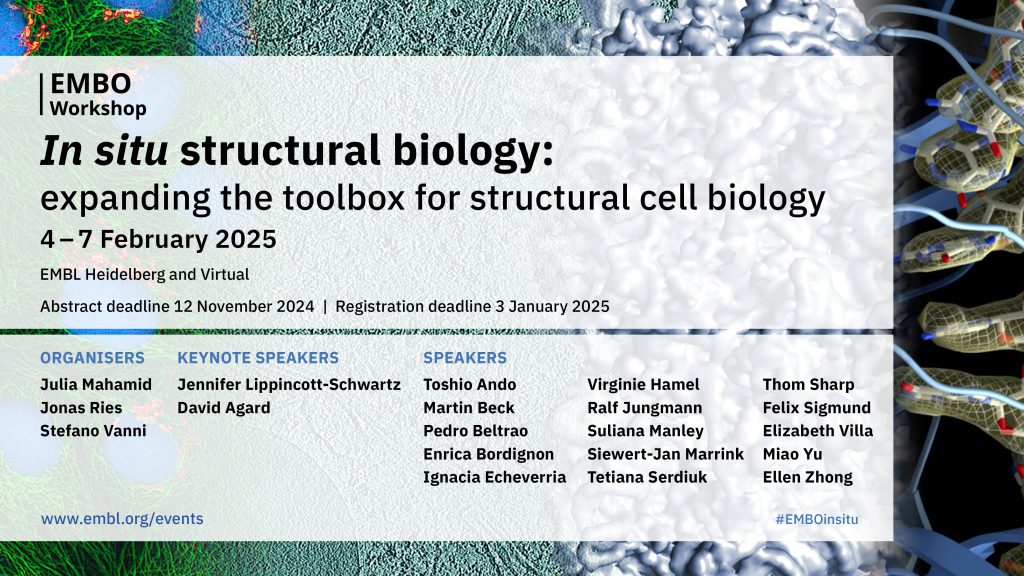
Five takeaways from ‘In situ structural biology: expanding the toolbox for structural cell biology’
Written by Ahmed Adel Ezat, PhD, lecturer at the Biophysics Department, Faculty of Science Cairo University, Egypt.
ℹ️ Ahmed attended this EMBO Workshop virtually, which is a great opportunity for those who are unable or unwilling to travel. Find out more about our event platform and the benefits or virtual participation and what it’s like to participate in our meetings remotely by having a look at another one of our blog posts – perhaps that’s a good option for next time you want to join us but aren’t able to make it all the way to Heidelberg!
Now, back to the #EMBOinsitu report:
In 1958, John Kendrew reported the first X-ray structure of myoglobin at a low resolution of about 6 Å, marking the birth of the structural biology field. The scientific community acknowledged this breakthrough in 1962 when John Kendrew, along with Max Perutz, was awarded the Nobel Prize in Chemistry.
In 1985, Kurt Wüthrich reported the first NMR protein structure. His contributions further advanced the field of structural biology and were recognised with the Nobel Prize in Chemistry in 2002. The peak advancements in structural biology have been achieved following the development of cryo-electron microscopy (CryoEM) for solving biomacromolecular structures. In 2017, three scientists—Jacques Dubochet, Joachim Frank, and Richard Henderson—were awarded the Nobel Prize in Chemistry “for developing cryo-electron microscopy for the high-resolution structure determination of biomolecules in solution.”
Afterwards, CryoEM methods have continued to advance, and new workflows and pipelines have been proposed to tackle specific scientific questions. CryoEM and cryo-electron tomography (CryoET) have matured structural biology and facilitated the study of large macromolecular assemblies and molecular machines in their native cellular context. These advancements are helping build a cell structure atlas detailing the anatomy and morphology of cellular content at near-atomic resolution.
Briefly, I will highlight the five key messages from this four-day meeting:
1. Cellular processes are defined by complex networks of biomolecular interactions.
The structure–function relationship relates the biomolecular function to structure, which encodes structural dynamics, and its interactions with other cellular partners modulate its function. It was difficult to harness such complex interactions between different parts of higher macromolecular assemblies at near-atomic resolutions, but thanks to advancements in Cryo(EM/ET), these have largely enabled us to solve these complex structures to understand the precise mechanism of function. Several talks showcased how in situ approaches monitor macromolecular assemblies to give more detailed mechanistic understanding of these cellular processes, from apoptosis, autophagy, and protein translation to motile cilia assembly and biomolecular condensate formation.
2. The new era of structural cell biology is in situ.
In vitro structural cell biology is the past, and in situ structural cell biology is the present and future of our mechanistic understanding of different cellular processes. We should observe biomolecular entities within their cellular context to fully grasp their interactions and functions. To achieve these goals, scientists have developed several workflows and pipelines to observe supercomplexes inside intact cells. Lea Dietrich, from the Max Planck Institute for Brain Research, Germany, showed the structure and arrangement of the mitochondrial oxidative phosphorylation machinery using cryo-lamella focused ion beam (FIB) milling combined with subtomogram averaging.
Richard Stefl, CEITEC Masaryk University, Czech Republic, showed the mesoscale organisation of transcription biomolecular condensates that are difficult to observe due to their dynamic nature and small size. He combined single-particle cryo-EM/ET with coarse-grained simulations to study the molecular structure of a condensate containing the phosphorylated RNAPII elongation complex and the elongation factor RECQ5.
3. Integration is changing the game.
There is no method without any limitations, and CryoEM/ET cannot reach high resolutions in situ due to physical constraints within the cell and the very crowded nature of the cellular milieu. Integration of data from multiple techniques is essential across all scales and builds a more finely detailed view of the macromolecular system on both spatial and temporal scales. Till Rudack, University of Regensburg, Germany, presented an integrative computational modelling strategy that combines both structural bioinformatics methods with data from structure-resolving experiments to investigate the functional cycle of molecular machines, and applied it to study ATP-hydrolysis-driven protein recycling by the proteasome and RNA degradation by the exosome. Altair Chinchilla Hernandez, Pompeu Fabra University, Spain, used the Integrative Modelling Platform (IMP) to build a model of the exocyst by combining nano-precise measurement of distances between fluorophores in situ and AlphaFold model predictions. Caitlyn McCafferty, University of Basel, Switzerland, showed the integration of cryo-ET with AI structure prediction, cross-linking mass spectrometry (XL/MS), and ultrastructure expansion microscopy (U-ExM) to explore ciliary structures and assembly.
4. Advanced imaging innovations are pushing the boundaries of structural cell biology.
Super-resolution microscopy provides precise single-molecule localisation of macromolecules in live cells. When combined with CryoEM/ET, they provide accurate single-molecule localisation and near-atomic resolved structure in the native physiological state. Several talks presented different workflows to combine the best of both methods in order to expand the toolbox of structural biology.
Ralf Jungmann, Max Planck Institute of Biochemistry, Germany, showed the use of DNA origami as barcodes to label different molecules in the cell at single-molecule resolution to acquire high resolution. Nikolai Klena, Human Technopole, Italy, presented cryo-ExCLEM, a method to perform ultrastructural expansion microscopy (U-ExM) on cryo-ET samples. This facilitates super-resolution correlation with cryo-ET structural information that improves protein localisation in native cellular environments.
Peter Dahlberg, SLAC National Accelerator Laboratory, USA, introduced a tri-coincident system that integrates light, ion, and electron microscopy at a single focal point in order to guide the milling process in a registration-free manner.
Toshio Ando, Kanazawa University, Japan, presented the power of high-speed atomic force microscopy (HS-AFM) in resolving the dynamics of macromolecules in near-physiological conditions.
Gaia Perone, Human Technopole, Italy, presented a cryo-lift-out procedure, SOLIST (Serialised On-grid Lift-In Sectioning for Tomography). This method was developed to study liquid-like biomolecular condensates.
These developments have expanded the tools to be applied beyond supercomplexes to organelles and compartments.
Suliana Manley, EPFL, Switzerland, presented the combination of cryo-ET, live-cell phase and super-resolution fluorescence microscopy to dissect the bridges between mitochondria and neighbouring organelles.
Jana Kroll, Max Delbrück Center for Molecular Medicine, Germany, presented a workflow for time-resolved in situ cryo-ET, combined with biosensors, optogenetics, and plunge freezing, to morphometrically and biophysically characterise the individual states of vesicle fusion in neurons under near-native conditions.
5. Computational modelling is at the forefront of structural biology.
The combination of diverse information from multiple experimental techniques and computational data aids in the modelling of large supercomplexes of the cell. Thus, computational data integration is revolutionising structural cell biology and mapping the entire cell at different spatial and temporal scales. It adds new molecular, kinetic, and dynamical details to our description of cellular processes.
Siewert Jan Marrink, University of Groningen, The Netherlands, employed the coarse-grained Martini model to construct a dynamical 3D model of the whole cell.
Margot Riggi, Max Planck Institute of Biochemistry, Germany, presented agent-based modelling (ABM) as a workflow to build intuitive, 3D simulations that explicitly include intramolecular conformational changes in addition to Brownian diffusion and molecular interactions.
Frosina Stojanovska, EMBL Heidelberg, Germany, presented CryoSiam, a self-supervised deep learning framework for detailed voxel-level and subtomogram-level segmentation and classification of cryo-ET data. It effectively distinguishes fine structural details through voxel-level segmentation and maps subtomograms into an embedding space that separates distinct macromolecular complexes.

The EMBO Workshop ‘In situ structural biology: expanding the toolbox for structural cell biology’ took place between 4 – 7 February 2025 in Heidelberg, Germany.
Read also about the poster prize winners from ‘In situ structural biology: expanding the toolbox for structural cell biology’ and find out more about their research in another blog post from the meeting.
Did you know that you can become an event reporter and receive a conference fee waiver in exchange? Find out how to do that by visiting our Become an event reporter page.