‘Building networks: engineering in vascular biology’ – check out the awarded posters!
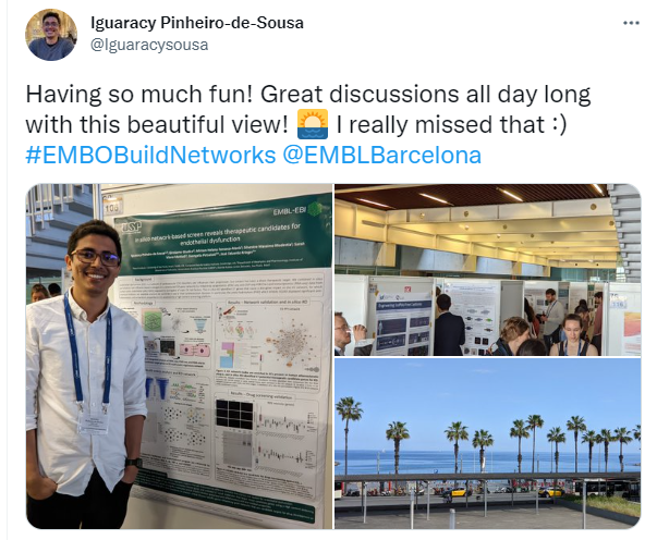
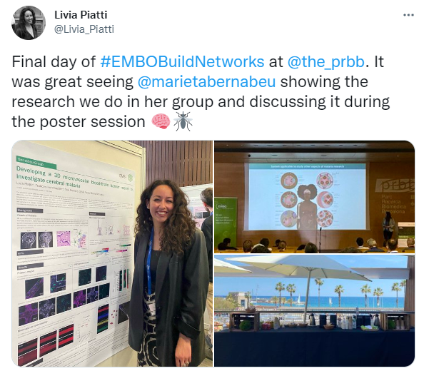
There were 5 poster prizes awarded at the recent EMBO Workshop ‘Building networks: engineering in vascular biology’, hosted by EMBL Barcelona at the Barcelona Biomedical Research Park (PRBB) and bringing together vascular biologists and bioengineering researchers from across Europe and beyond, who are pioneering new tools towards understanding vascular biology in health and disease. For many participants this was the first on-site meeting that they attended since early 2020 and the spirits were high during the three days full of exciting science, exchanging ideas, presenting latest research, catching up with old friends and making new ones.
We also didn’t have any complaints regarding outdoor coffee breaks on a sunlit deck overlooking the sea! There were two live poster sessions during which the presenters could discuss their research (over snacks and drinks!) – their work was then voted for by other attendees and speakers. We are pleased to be able to share with you the research from the five winners of the best poster prizes: congratulations to Claire, Akinola, Irene, Nensi and Anjali!
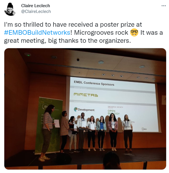
Contact guidance of vascular endothelial cells on microgrooved substrates: influence of groove dimensions and cell density
Presenter: Claire Leclech

Abstract
In healthy arteries, endothelial cells (ECs) exhibit different morphologies: elongated and aligned in the direction of blood flow or more cuboidal in regions of arterial branches and bifurcations. Factors that regulate EC morphology and alignment are of interest, particularly in light of the observation that atherosclerotic lesions preferentially form in regions where ECs are less aligned and elongated. In vivo, the basement membrane to which ECs adhere is a patterned and topographic surface. We are interested in how this substrate topography may regulate EC shape and alignment and are exploring these questions in vitro using microfabricated surfaces.
When cultured on substrates composed of parallel arrays of microgrooves, ECs align and elongate in the direction of the grooves, a process called contact guidance. We show that we can control the extent of this contact guidance by modulating the groove dimensions (spacing, width, and depth). In particular, we demonstrate that increasing groove depth (from 1 to 6 μm) leads to the most pronounced cell elongation and alignment. We also investigate the influence of cell density on the response to microgrooves by comparing the response of individual cells to monolayers of low or high density. Interestingly, we observe progressive loss of cell alignment and elongation on microgrooves for increasing cell density/culture time, associated with remodeling of the actin cytoskeleton and focal adhesions (FAs).
We are investigating the mechanisms underlying this depth- and density-dependent response of ECs to the microgrooves and propose that a competition between cell-substrate and cell-cell adhesion may explain the existence of different mechanisms. In individual cells, the depth-dependent response predominates, driven by FA clustering and protrusion dynamics, while in highly confluent monolayers, ECs respond primarily to the secreted basement membrane and lose the response to substrate topography.
Beyond highlighting fundamental mechanisms of shape modulation and contact guidance in ECs, the results of this study can also prove useful in the field of implantable endovascular devices where surface topographic functionalization may constitute a promising strategy for improving device efficacy.
Engineering 3D vascularized cardiac microtissues on-chip
Presenter: Akinola Akinbote

Abstract
Coronary microvessels are implicated in many cardiovascular diseases (CVDs) and their dysfunction is associated with adverse clinical outcomes. These outcomes vary by biological sex and are hypothesized to differ based on estrogen’s cardioprotective effects. However, endothelial-dependent contributions to CVDs and sex-based differences are still largely unexplored, in part due to inadequate models of the cardiac microvasculature. The advent of iPSC-derived Cardiomyocytes (CMs) has resulted in a growth in cardiac models. 3-dimensional cultures and co-cultures with non-myocyte populations, such as cardiac fibroblasts and endothelial cells (ECs), have also been shown to improve cardiomyocyte maturation in vitro (marked by improved calcium handling and metabolic maturation); yet the impact of beating cardiomyocytes on cardiac microvascular remodelling and barrier function is not well understood. By employing microfluidic models of microvessels and iPSC-derived cardiac spheroids we can explore these complex CM-non-myocyte interactions in a controlled and quantifiable manner. We are generating adult-derived cardiac microvessels integrated with cardiac organoids to reveal the contribution of 1) non-myocytes (cardiac fibroblasts and endothelium) to cardiomyocyte function and 2). the effect of beating cardiac organoids on local microvascular remodelling. This unique vascularized cardiac model will be useful for understanding complex vascular-myocyte interactions and may provide clues to the role of sex hormones in promoting both endothelial and cardiac function.
Due to the confidentiality of the unpublished data, we cannot share the poster.
Temporal adaptation of vascular patterning
Presenter: Irene M. Aspalter

Abstract
Sprouting angiogenesis is highly dependent on effective decision making between endothelial cells (ECs). The feedback between Vegf/Dll4/Notch is well established during the collective selection of tip cells of new vessel sprouts.
Our prior work demonstrates that additional signals (e.g. sema3E-plexinD1) can alter the tip cell selection speed by acting as time-keepers during Dll4/Notch signalling. Changes to the timing of this process alters the vascular network density.
It was believed that tip cells form filopodia post selection, aiding the migration towards Vegf. However, our recent in silico models, validated in vivo, show that ECs form filopodia first, irrespectively of whether they are selected as tip. Our simulations predict that filopodia speed up tip cell selection by moving Vegf-receptors towards the Vegf source, creating a sensory-motor-feedback that speeds up Dll4 production. Indeed, my preliminary in vitro data shows Vegf-receptors at the tip of filopodia. This suggests a vital role of filopodia as time-keepers during tip cell selection.
We aim to better understand the role of filopodia and other time-keepers to fine tune vascular patterning and network topologies.
Using microcontact printing I am developing a method to closely investigate the tip cell selection timing while modulating the involvement of filopodia and other pathways. Thin printed lines of extra cellular matrix allow ECs to interact, but not to swap positions, prohibiting disruption of Notch patterns in order to observe when stable patterns establish.
pERK has been previously shown as suitable tip cell marker in zebrafish and is also a useful marker in my system. Using pERK as readout, my preliminary data shows different selection patterns of tip/stalk cells in the presence or absence of Notch inhibitors, and we are currently developing an analysis pipeline for robust quantification.
This system will be used to carefully dissect the mechanism by which filopodia influence tip/stalk cell selection, with the help of molecular manipulation (growth factors/inhibitors) and micro manipulation (photo-activatable probes).
Our work will shed new light on the tip cell selection process and will offer new targets for therapeutic approaches targeting temporal regulation of vascular patterning, network topology and branching density.
Resolving vascular endothelial junctions with correlative fluorescent light microscopy and cryo-electron tomography (cryo-ET)
Presenter: Nensi Alivodej

Abstract
Endothelial cells (ECs) form the inner lining of blood vessels, where they adhere to one another via junctional complexes, namely adherens and tight junctions, to regulate the integrity and permeability of the vascular barrier. These junctions are critical for tissue development and homeostasis and are structurally different across organs. For example, ECs of the brain possess strong tight junctions that are central to the formation of the blood-brain-barrier while ECs of the lung rely on adherens junctions to maintain vascular permeability at the interface of the blood-air barrier.
While the molecular composition of adherens and tight junctions is well studied, less is known about the assembly of junctional proteins in their native environment and their interaction with the cytoskeleton. Past cryo-electron tomography (cryo-ET) studies have been unsuccessful in resolving the structure of these junctions in situ owing to the difficulty in identifying them under cryo-electron microscopy. To circumvent this limitation, we have established a pipeline employing state-of-the-art correlative fluorescent light microscopy and cryo-ET in order to resolve junctions in ECs. We have isolated ECs from different organs, including the lung, brain and aorta, in mice expressing an EGFP-tagged VE-cadherin knock-in protein and have successfully grown them on electron microscopy (EM) grids. After cryo-fixation via plunge-freezing, we identified areas of interest expressing the fluorescently-tagged VE-cadherin using light microscopy and correlated those areas to EM images for localizing junctions. The areas were then processed to obtain a series of transmission EM images at multiple angles in order to reconstruct the three-dimensional organization of the junctions. This pipeline has allowed us to obtain the very first EM images of in situ adherens junctions from wholly preserved lung ECs, where individual VE-cadherin proteins, auxiliary adapter proteins and underlying cytoskeleton can be visualized with a pixel size of 0.22 nm. We are optimizing our workflow to also resolve tight junctions of the blood-brain-barrier. Altogether, we have established a scalable pipeline to characterize and compare the tissue-specific structural organization of vascular junctions with unprecedented sub-nanometer resolution.
Due to the confidentiality of the unpublished data, we cannot share the poster.
Geometry of self-assembled DNA nanostructures influences in-vitro angiogenesis in HUVECs
Presenter: Anjali Rajwar

Abstract
DNA nanotechnology involves fabricating small strands of DNA to design nano-objects in 1D, 2D and 3D with precise control of shape and size that have been utilized in many applications.
DNA nanostructures have been investigated for their ability to influence cellular behaviour and functions. Recently, new emergent functionalities of DNA nanodevices as a class of biomaterials with immense capacity to interface with biological systems and vast potential in disease diagnosis and therapeutics have emerged. DNA nanostructures, which are chemically robust and biocompatible in nature, have been surface modified and structurally fine-tuned to find emerging applications in stem cell therapy and tissue regeneration. DNA nanostructures can be used for therapeutic angiogenesis, which involves the formation of new blood vessels, and can be used to treat ischemic diseases such as stroke or heart failure. This study looks at how the structural topology of DNA nanostructures affects their ability to stimulate endothelial cell angiogenesis.
We examined the potential of four different DNA nanostructure geometries on the differentiation of human umbilical vein endothelial cells (HUVECs). While different DNA nanostructure geometries successfully induced angiogenesis and cell migration in HUVECs, tetrahedral DNA cages demonstrated the greatest uptake and angiogenesis potential, indicating that not only the composition of materials, but also the 3D arrangement of ligands may play a role in stimulating the angiogenesis process.
Taken together, this research can lay the groundwork for future studies involving DNA nanocages for biological and biomedical applications, explicitly applying their surface topologies in bioimaging, drug delivery, immune activation, and tissue engineering.
Congratulations to all five winners!
The EMBO Workshop ‘Building networks: engineering in vascular biology’ took place from 9 – 11 May 2022 at EMBL Barcelona.