Generating meaningful images – a report from Seeing is Believing 2019
By event reporters Liz Haynes @actin_crazy and Stephan Daetwyler @Daetwyler_St
The field of biology owes some of its most compelling discoveries to careful visual observation. From Van Leeuwenhoek’s use of new microscopes to describe microscopic “animalcules” in the late 1600s, to Ramon y Cajal’s pioneering 19th century work illustrating beautiful and complex neuronal architecture. Images inspire us, help us generate new hypotheses, and shed light into the tiny worlds yet unexplored. Indeed, these observations uniquely help us understand the structures and dynamics of life, something that would not be achievable with approaches like biochemistry alone.
The images are only as valuable as the amount of information that we can deduce from it.
Generating meaningful images, however, is not an easy task. There have always been limits to what we can observe, due to the properties of the sample or the techniques that we can apply to it. These are the boundaries that microscopists seek to push. A successful imaging experiment requires an amenable sample, a contrast agent to reveal the structures of interest, and a microscope that is capable of capturing an image at a relevant scale. Moreover, the images are only as valuable as the amount of information that we can deduce from it. Therefore, image storage, accessibility and analysis are crucial. Each one of these steps offers opportunities for optimisation and new technologies.
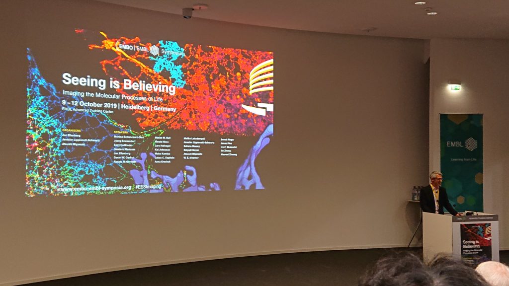
The EMBO | EMBL Symposium “Seeing is Believing: Imaging the Molecular Processes of Life” (9-12 October 2019) presented us with exciting new developments in all of these fields, coupled with a drive to make new progress available as quickly as possible to the community through preprints, open-source initiatives, and resource sharing.
Advances in sample preparation
At the heart of every imaging approach is the sample. Even the best microscope is ineffective with dim or improperly prepared samples. At Seeing is Believing, we saw an emphasis on using expansion of samples to help overcome the resolution limits of microscopy and solve some traditionally difficult problems. In particular, we were impressed with expansion-based approaches to study centriole structure (Paul Guichard, Ultrastructural Expansion Microscopy) and resolve microtubules tightly packed within axons (Lukas C. Kapitein). By far, the biggest emphasis in sample improvement was on the development of new fluorescent probes and biosensors. Kai Johnsson presented design strategies for the improvement of live cell dyes, and introduced new MaP dyes that are SNAP and HALO compatible, and importantly require no wash to clear unbound probe. Periklis Pantazis presented a mechanosensor based on the Piezo1 stretch activated ion channel, allowing users to visualise mechanical stress within a live cell. Atsushi Miyawaki wowed the audience by meeting the challenge to “be better than a firefly” with a new variant of luciferase named AkaBLI, which his lab generated through targeted evolution. This improved luciferase allowed them to visualise neuronal activity within freely behaving mice and marmosets.
Advances in microscopy
The features of our microscopes directly determine which questions we can address. Seeing is Believing highlighted exciting new development in building cutting-edge microscopy tools. Reto Fiolka presented a novel single-objective light-sheet microscope enabling imaging of live cells in microfluidics devices or 3D environments with 200 nm lateral resolution. Kevin Dean complemented novel light-sheet development by presenting an axially swept light-sheet microscope ideally suited for all clearing techniques that provides an unprecedented field of view enabling whole tissue imaging with sub-micron resolution. With her imaging approach, Alexandra Pacureanu surprised the audience with how X-ray holographic nano-tomography is capable of resolving the fine, dense and complex neuronal circuitry in large tissues or even organism providing a new route to understand how the nervous system processes information.
Further impressive advances were presented in fast volumetric imaging (Lars Hufnagel, light field imaging) and high-resolution imaging, e.g. MINFLUX by Stefan Hell, correlative EM imaging by Harald Hess and Lucy Collinson, GI-SIM/LLS-SIM by Dong Li, and 3D-STED deep in a tissue by Joerg Bewersdorf.
Advances in data analysis
All acquired data is meaningless if we cannot extract information from it. At Seeing is Believing, it became obvious how artificial neuronal networks have become important for image analysis. Applications range from segmentation to denoising an image (BGnet, W.E. Moerner and Noise2Void, A. Krull/Florian Jug). Particularly, the convolutional network architecture U-Net has become an important tool. To provide a user-friendly environment to apply those state-of-the art image analysis tools, Anna Kreshuk presented the iLastik platform as an easy to use tool. A new fundamental approach to handle, visualise and process the large amount of data coming from the microscopes was presented by Ivo Sbalzarini. Instead of using pixels to save an image, adaptive particles approximate the image content. Furthermore, Gaudenz Danuser gave a thought-provoking talk on how current perturbation-based approaches in cell biology can mislead us in our analysis. Danuser emphasised that the observed phenotype from a perturbation of a system (e.g. loss of a protein’s function) is not equal to the real function of the gene. For example, cutting a wire from the battery to the electronic board of radio would lead to the “phenotype” loss of sound. However, the function of the wire was simply to provide power to the radio, not to produce sound! As a better perturbation-free alternative, Danuser introduced a concept used in econometrics known as Granger causality.
Advances in biology
All of these new developments culminated in impressive new insights into biological processes. There were many talks on mitochondria and endoplasmic reticulum dynamics revealed by novel live-cell super-resolution techniques. Suliana Manley gave one of the most intriguing of those talks, on modes of asymmetric and symmetric mitochondrial division.

Jennifer Lippincott-Schwartz also gave a stunning presentation on how RNA granules can hitch a ride through an ANXA-11 mediated connection to lysosomes, and how ALS associated mutations in ANXA-11 break this connection. Furthermore, an intriguing new mRNA reading frame sensor (Moon and Sun tags) was presented by Sanne Boersma of the Tanenbaum lab to understand stochasticity of mRNA translation.
To conclude, the field of microscopy has grown so much that some may feel we have solved all the theoretical problems, and only engineering challenges are left – hardware improvements, new materials, new engineering solutions. At the closing dinner of the conference, however, Atsushi Miyawaki from RIKEN beautifully summarised how he felt about the future of microscopy, and of Seeing is Believing. Standing in the banquet hall of the Heidelberg Castle, he told us that castles in Japan remain unfinished. This state of incompletion is not due to any fault of the architects, but a feature of beauty, as it was believed that things that were incomplete had room to grow, and that growth is valuable. No matter how high our achievements are in the field of microscopy and image analysis, there will always be unforeseen avenues of growth. Attending Seeing is Believing has hopefully prepared us to follow those avenues, and to share what we find so we may all grow together.
For a more comprehensive summary of all talks presented at Seeing is Believing, and to get links to preprints, publications, and resources, visit our blog at https://seeingbelievingweb.wordpress.com/