Artificial intelligence revolutionises microscopy
From using artificial neural nets to elevate biological image analysis to applying machine learning to probe organoid cultures, AI-associated techniques contributed significantly to EMBL’s microscopy-based research activities.
Artificial intelligence (AI) proved to be a game-changer in the fields of microscopy and image analysis in 2021.
AI can help overcome the limitations of traditional tools. For example, light-sheet microscopy and light-field microscopy hold different advantages and challenges. To take advantage of the benefits of each technique and with the help of strong GPU compute power provided internally by EMBL IT Services, researchers at EMBL developed a machine-learning approach that uses light-field microscopy to image large 3D samples and light-sheet microscopy to train AI algorithms, which then create an accurate 3D picture of the sample.
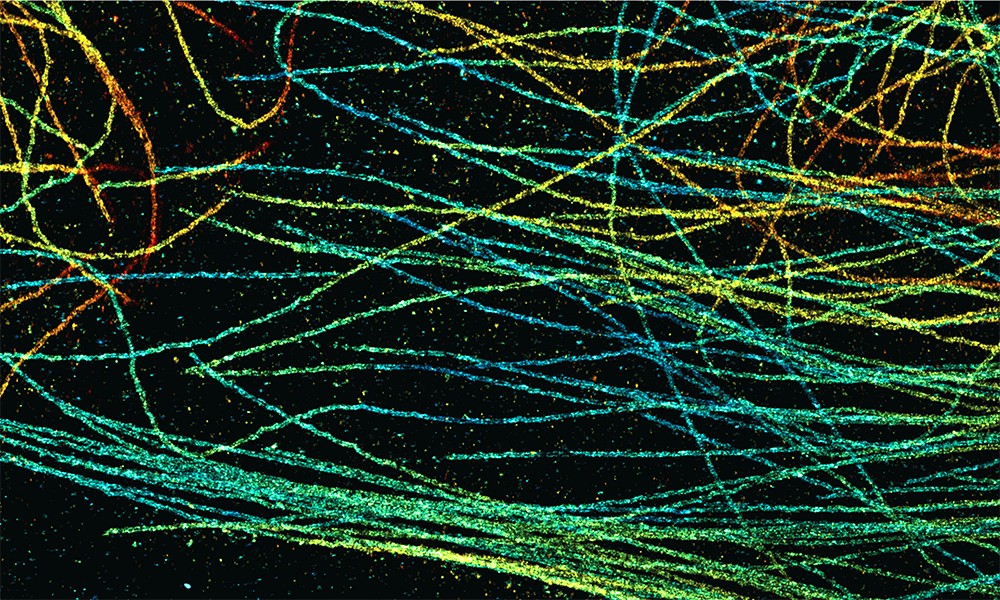
Similarly, the field of super-resolution microscopy also benefited from an injection of AI-driven innovations. “One of the biggest limitations of super-resolution microscopy is how long it takes to collect data from the microscope,” said Lucas-Raphael Müller, former master’s degree student in the Ries Group at EMBL Heidelberg.
To overcome this challenge, EMBL scientists and collaborators developed DECODE (DEep COntext DEpendent), an open-source computer program based on a neural network that learns from training data. Using this, imaging speeds can be increased up to tenfold with minimal loss of resolution.
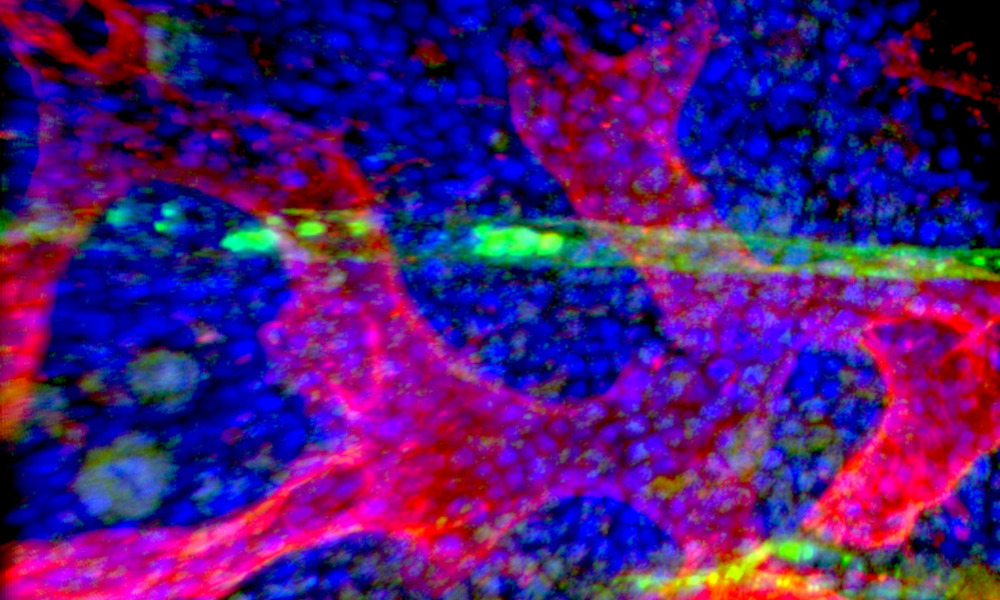
At EMBL Barcelona, researchers developed MOrgAna, an open-source software that uses machine learning to analyse images of organoids and perform segmentation. Due to its ability to capture the complex morphological features of organoids, MOrgAna performs better segmentation than previously existing platforms.
EMBL research results in this area represent the future of science: open, collaborative, interdisciplinary, and designed to accelerate knowledge transfer.
“The rapid progress in adopting deep learning/AI in microscopy and image analysis never fails to amaze me, as such platforms enable us to glean trailblazing insights from mammoth amounts of data or imagery, quickly.”
— Jia Le Lim, PhD student, Trivedi Group at EMBL Barcelona, whose response to a pandemic that limited her lab access in 2021 was to help create a ‘smart’, easy-to-use, open-source software, MOrgAna, for analysing organoid images in a high-throughput manner.
Explore more 2021 EMBL AI highlights:
References
Wagner N et al. (2021). Deep learning-enhanced light-field imaging with continuous validation. Nature Methods, 7 May 2021. DOI: 10.1038/s41592-021-01136-0
Speiser A, Müller LR et al. (2021). Deep learning enables fast and dense single-molecule localization with high accuracy. Nature Methods, 03 September 2021. DOI: 10.1038/s41592-021-01236-x
Gritti N, Lim JL et al. (2021). MOrgAna: accessible quantitative analysis of organoids with machine learning. Development, 8 September 2021. DOI: 10.1242/dev.199611