
A flip book for biological systems
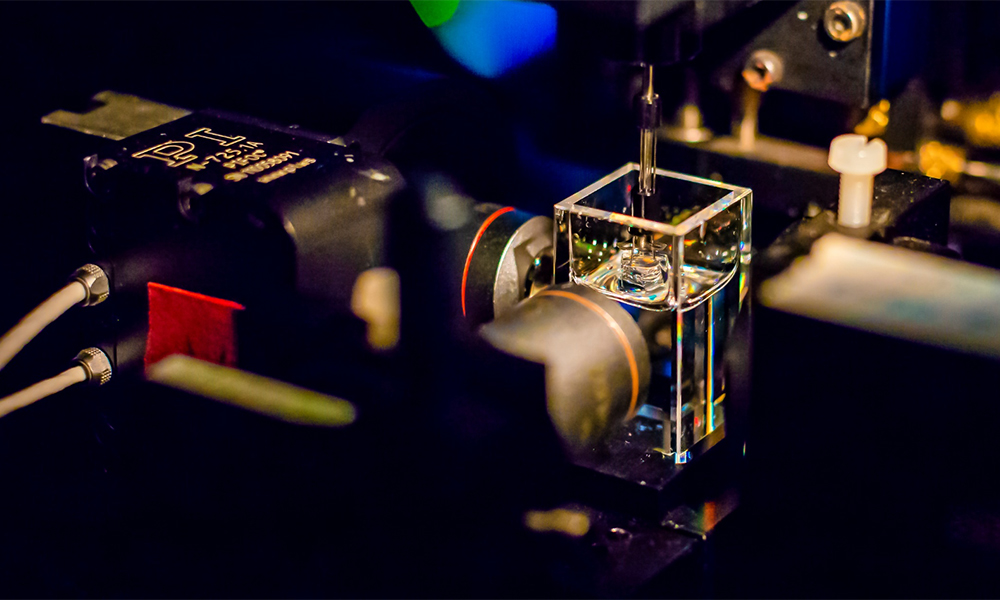
In the Mesoscopic Imaging Facility (MIF) at EMBL Barcelona, researchers study the details of biological systems in the context of organs, body parts, or entire organisms. This image shows OPTiSPIM1, one of the custom light-sheet microscope setups available at the facility.
Light-sheet microscopy produces images of biological samples one thin slice at a time. This minimises photodamage caused by the excitation radiation used to illuminate the sample. The sample emits fluorescence, which is collected for each slice by the detection optics. At the end of this process, the light-sheet images obtained along the entire depth of the sample are stacked on top of one another. You can visualise them as if you were leafing through a flip book.
An example of a stack of images ‘flipping’ through the depth of an ovary. The video shows a mouse ovary and oocytes, or egg cells, at various stages of maturity. Their dimensions range from 10 to 50 micrometres. Credit: Arturo D’Angelo/CRG; Montserrat Coll Lladó/EMBL Barcelona
To be observed using OPTiSPIM1, the biological sample is encased in agarose and suspended in a transparent chamber of clearing medium (visible in the image). The clearing medium makes the sample transparent and allows light to pass through with minimal scattering. The chamber is placed at the intersection of the illumination and the detection axes. This setup enables researchers to achieve high-resolution imaging in thick samples that are opaque in their natural state.
The MIF are world-leading experts in this technique – indeed Jim Swoger, head of this facility, is one of the original inventors of light-sheet imaging, back in 2004, while he was in Ernst Stelzer’s Light Microscopy group.
If you have a stunning picture of your science, your lab or your site, you can submit it here.


