
A loopy baseline
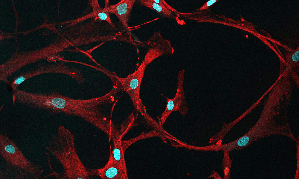
Studying cancers means also knowing what healthy cells look like. In this case, mesenchymal stromal cells (MSCs) from healthy bone marrow are a bit ‘loopy’.
This beautiful image comes from a confocal microscope, which captured the CD90 protein (shown in red) that marks the surface of these cells. Researchers from EMBL’s Zaugg group study leukaemia onset and were looking to see whether CD90 would be an effective marker to identify MSCs in bone marrow.
Targeting the microenvironment of leukaemic cells, in this case MSCs, can result in novel leukaemia therapies. Images like this can serve as a baseline for further research to study them in the context of leukaemia.
Leukaemia is a cancer of the blood and bone marrow, and these scientists are studying the most prevalent types of leukaemia that tend to strike between the ages of 65 and 74.
Credit: Anna Mathioudaki/EMBL
If you have a stunning picture of your science, your lab or your site, you can submit it here.


