
Gazing at healing wounds
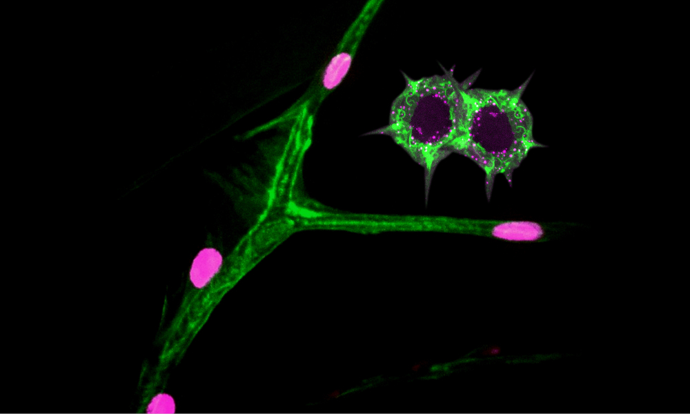
What may look like a branch of a tree with the first flower buds emerging after winter are, in fact, tracheal cells of a fruit fly larva and their nuclei. For decades, scientists have been using the fruit fly Drosophila to understand biological processes; 60% of fruit fly genes have been conserved throughout evolution and are also present in humans.
Former EMBL Heidelberg researchers Parisa Kakanj, together with colleagues from the Institute for Genetics of the University of Cologne, the Max Planck Institute for Biology of Ageing, and the CECAD and CMMC in Cologne, developed a new method to study molecular interactions in living fruit fly larvae.
Parisa then used this method to study wound healing. To create this particular image visual artist Mona Kakanj, who is also Parisa’s sister, used a snapshot from a time-lapse video Parisa created for her study on Drosophila larvae and edited it in Photoshop.
In the image the plasma membrane and cytoplasm of the larval tracheal cells appear in green and the nuclei appear in magenta. The two star-shaped objects in green are the plasma membranes of cells surrounding an epidermal wound.
Credit: Parisa Kakanj & Mona Kakanj/EMBL
If you have a stunning picture of your science, your lab or your site, you can submit it here.


