
Mapping space inside the cell
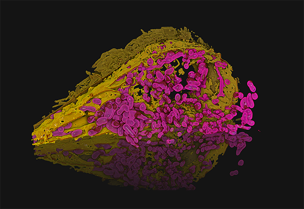
In this image, Julian Hennies from the Schwab Team has reconstructed the 3D structure of a human cell’s organelles. The open cell reveals the mitochondria (pink), the endoplasmic reticulum (yellow), and parts of the plasma membrane (khaki). Although the nucleus is missing in this image, you can make out its location – the void on the right-hand side, around which the organelles seem to organise.
The endoplasmic reticulum is a network of membranes involved in the manufacture and transport of proteins inside the cell. Hennies is studying its morphology and its points of contact with other organelles. For instance, the dark red spots on the mitochondria are where they meet the endoplasmic reticulum. To visualise these intimate contacts in their spatial context, Hennies had to combine multiple techniques, including focused ion beam scanning electron microscopy (FIB-SEM) and image segmentation.
When applied to a stack of images, Hennies’ method makes it possible to reconstruct an object in 3D, but it is no straightforward procedure. He had to automate the segmentation of the images in his colossal datasets using a machine learning approach, to avoid spending all of his doctoral studies running the analysis manually.

If you have a stunning picture of your science, your lab, or your site, you can submit it here.


