
Inside out, and grub becomes fly
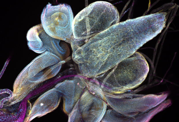
The three bluish blobs shown in the top right corner of this image may not resemble the sphere of noodles that is the human brain, but they are still essential – at least for the fruit fly. This Picture of the Week shows the brain lobes of Drosophila. It’s an insect so tiny and so common that you may forget its importance for research when observing it hover over ripe fruit in the summer.
Here, Daniel Rios dissected a fruit fly larva to stain specific proteins in its internal organs with fluorescent antibodies (blue and yellow; cell nuclei appear in pink). Organs like the brain lobes, but not only these. The other structures in this picture are organs that start developing in the larva and change tremendously during metamorphosis to form the future wings and legs of the adult fly.
Just like a caterpillar turns into a butterfly, a grub rearranges its anatomy to become a fly. Shaping new tissues and complex organs like wings is the work of many proteins produced by the protein factories of the cell – two organelles called the endoplasmic reticulum and the Golgi apparatus. Taken with a confocal microscope, this picture shows the distribution of the endoplasmic reticulum within different tissues in a colourful overview.

Credit: Daniel Rios/EMBL
If you have a stunning picture of your science, your lab or your site, you can submit it here.


