Five decades of EMBL visits
Structural biologist Shlomo Trachtenberg, who has made research trips to EMBL from Israel since the late 1970s, reflects on the boost EMBL’s technology provided his research, the ingredients for an ideal research institution, and his ongoing awe of microscopy.
It wasn’t long after the first labs at EMBL Heidelberg were constructed that a young scientist from Israel was thrilled to come for a short visit. He looked forward to making use of equipment that significantly surpassed that at his home institution and allowed him to exploit a fledgling tool known as cryo-electron microscopy.
After that first visit, Shlomo Trachtenberg began what would become near-yearly trips to Heidelberg, spending time with many EMBL group leaders and their labs throughout the years: Kevin Leonard, Jacques Dubochet, Marc Adrian, Gareth Griffiths, Max Haider, Willem Tichelaar, Andy Hoenger, Ernst Stelzer and now Simone Mattei at the Imaging Center. And while there might have been sharp disparity between available microscopy between the two countries, the intellectual exchange between Trachtenberg and EMBL scientists was on far more equal footing, creating a two-way information exchange that helped everyone’s research endeavours.
Trachtenberg has spent his research career at Hebrew University of Jerusalem, where he is now a professor emeritus. Using primarily light and electron microscopy, he has spent most of his career working with bacterial flagella (thin, rotary helical propellers) and Spiroplasma (helical bacteria with internal linear motors). Trachtenberg has been interested in this bacteria’s remarkable motility and its underlying molecular machinery.
“Bacteria swim. And they swim quite fast,” he said. “They can swim 50-100 body lengths per second – the equivalent of a human being running 200 metres in a second. Notably, because they are small, the viscosity of them swimming in water is akin to us swimming in honey.”
As luck would have it, his own relationship with EMBL also extended to his daughter, Shira Trachtenberg, who spent three years at EMBL Heidelberg, starting as part of a student exchange programme after finishing architecture school. She was involved in erecting EMBL’s Advanced Training Centre (ATC) building.
“…EMBL was an accessible, cutting-edge, unique platform of science that I could not have found anywhere else. It was why I came here, and I enjoyed every second – personally and scientifically.”
Interestingly, Shira Tratchenberg was also involved in the erection of the heat-centre’s chimney, which is modelled after a seamed microtubule (from Andy Hoenger’s work). Shlomo Trachtenberg has been working on similar problems (non-helical perturbations) in bacterial flagella, which are now solved at atomic resolution. Most initial electron microscopy data of this project were collected at EMBL.
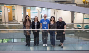
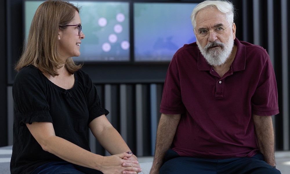
Rachel Mellwig, EMBL’s Electron Microscopy Facility Operations Manager, met Trachtenberg approximately 20 years ago when she was in her own first days at EMBL. In July 2023, when Trachtenberg had returned once again to EMBL for a month-long stint, she reached out to EMBL Communications. It was an opportunity to get an important perspective on EMBL through the years, especially as the institution approaches its 50th birthday in 2024.
“Everyone knew Shlomo, basically,” Mellwig said. “His experiences are a nice reminder that that is what the Imaging Centre is here for – to give access to these high-end machines, where oftentimes the manufacturer is onsite as well. EMBL is a place where these instruments are uniquely tweaked for the necessities of a variety of research happening here.”
We had the chance to sit down with Tratchenberg during his most recent visit to the EMBL Imaging Centre. Here are some of the key insights that emerged, in his own words:
Reflections on his science
I came to EMBL first as a student, because I wanted to study all the new techniques in microscopy. But later I realised that I couldn’t apply any of these techniques at my home University in Jerusalem.
We were working on the helical reconstruction of bacterial flagellar filaments using both STEM (scanning transmission electron microscope) and TEM (transmission electron microscope) and the combination of TEM and light microscopy.
When I went back as a professor to Jerusalem after experiences at NIH and then Brandeis University, I built from scratch a full-fledged cryo-electron microscopy and data analysis lab initially suitable for organismal to molecular studies. Unfortunately, something very common to university environments is to buy equipment but not bother to maintain it to the necessary high standards that researchers – myself included – require. This was my experience – an exasperating situation, to say the least. Worse, a common perception in many universities, as in mine, has been that electron microscopy is ‘service’ not ‘science’ and that cryo-EM is ‘not structural biology’. The unit I established has never functioned as intended. This is something young structural biologists aspiring to academic positions, of which there are not a few at EMBL, should be aware of and avoid at all cost.
That provided an even stronger incentive for me to come to EMBL to do what I couldn’t at home. Short-term fellowships or the status of a visiting scientist or visiting scholar allowed me to come once or twice a year. I came whenever I could because I could do all my high-end electron microscopy work here. I collected TEM and STEM data on bacterial and archaeal flagellar filaments and on linear motors of spiroplasmas. I analysed the image databases to study the symmetry and molecular structure of these motor polymers. EMBL was an accessible, cutting-edge, unique platform of science that I could not have found anywhere else. It was why I came here, and I enjoyed every second – personally and scientifically.
Reflections on early days at EMBL
What a great place to be in the ‘70s. I would work seven days a week. I collected data at EMBL, but processed the data at home. Microscopy – both light and EM – wasn’t available with this level of interaction. I think in my modest way, I contributed here too, sharing my own knowledge. It definitely wasn’t just a one-way relationship.
In the beginning, EMBL was basically a kind of free-flowing, relatively small, non-structured community. An important component of EMBL was the design and construction of electron microscopes, light microscopes, and cryo-EM hardware, which added flexibility, innovation excitement, and opportunities. All units were located in Heidelberg which created a rare concentration of knowledge, expertise, and unique people. Everything was open: open minded and open physically. As time passed, naturally it became more structured and geographically spread.
The marvel of microscopy and scientific discovery
The first time doing cryo-EM? It felt like a miracle.
At the time, there was nothing else like this microscope – Philips EM400 – on which most of the cryo-EM methodology and hardware were developed and pioneered. (Later on it would be the CM200FEG, a TEM). They have since been replaced, but I remember that initially to get an image, we went through many manual operations in a specific sequence. Now, it’s essentially the click of a button. And I was using technology advances developed at EMBL…Max Haider developed the liquid helium cooled STEM, which I used with Willem Tichelaar and Kevin Leonard (along with the VG cryo-HB5 STEM). We used these to study the helical symmetry and radial mass distribution of vitrified flagellar filaments. They really were exciting days.
When I recently bought a microscope for my granddaughter, it made me recall my own first impressions of an image. That too felt miraculous. Even today, that miracle isn’t gone for me.
I was also involved in the philosophy of science as a student, and I continue to be. When I first looked at bacterial motility and bacterial chemotaxis (how it moves toward helpful environmental conditions and/or away from repellent ones), I was exploring the way they are sensitive to their environment and respond to it. I thought about how that’s an experimental system of free-will studies, and that has never left me.
Scientists change through the generations. For example, two of the principal powerful microscope systems I worked with (highly important in the mass study of polymers, membranes, and single particles), the cryo-STEMs at EMBL and Brookhaven National Laboratory, simply vanished because people found them too complicated to use and stopped using them.
What Rachel is doing (thin sectioning of fixed- and resin-embedded specimens) is an opposite example. This kind of work was considered nearly obsolete, or outdated, in the high days of molecular cryo-EM, but it is undergoing a complete revival because of the advancement in technology and computing power. Whole brains and neural networks can be reconstructed at high resolution.
Favourite aspects of coming to EMBL
First, it was accessibility to cutting-edge hardware, technology, and interdisciplinary knowledge that wasn’t available elsewhere.
But there was also the freedom to concentrate purely on science and abstract thinking together with very concrete and technical ‘doing’. Coming from a very complicated and troubled part of the world, it was nice not to have the usual distractions — no TV, no newspapers, no news. I could wholly devote myself to my science.
I took advantage of the resources, and that included using free time to sit in the library at night or on weekends to think about things. I did a lot of writing.
There was always someone to discuss ideas with. It was rewarding to talk to people who shared my interests and expertise.
The future
It would be great to see EMBL provide a broader platform for things that can’t be done in a university environment in terms of cost, infrastructure, and maintenance. Going back to its roots while also moving forward. Many instruments are available commercially, but they are just instruments. EMBL is a place where instruments can be uniquely tweaked for high-end and non-standard necessities and applications.
Officially, I’m emeritus now, but I keep doing my science exactly the same way. I haven’t stopped working. And I will keep on coming to EMBL. I can’t think about my scientific work and its results without the presence of EMBL.