Timo Zimmermann
Team Leader
ORCID: 0000-0001-7098-0700
EditAdvanced Light Microscopy technology development and service provision
Team Leader
ORCID: 0000-0001-7098-0700
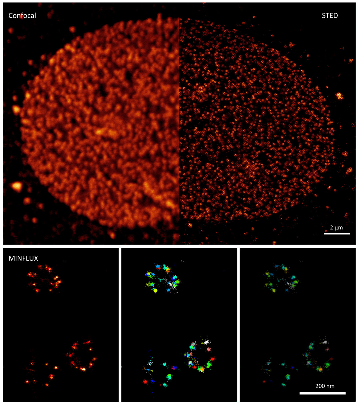
EditLight microscopy is a highly active and continuously developing technology field that has seen unprecedented progress in many of its areas in the past few years. These include the increase of resolution beyond the limitation of optical diffraction (super-resolution), the possibility of visualising larger and larger volumes at increasing speeds (by light-sheet imaging and nonlinear microscopy), and the possibility of extracting new forms of information from the sample (new labels, image modalities, and label-free imaging) at ever increasing sensitivity of detection.
To efficiently match the potential of such new imaging methods to the relevant questions of biological and medical researchers, imaging specialists are needed to interpret the biological question and the instrumentation and methods that can provide data to help answer it. This function is already performed very well in many light microscopy core facilities, but a gap can form between the service provision in core facilities and the leading edge of instrument and method development, because of the significant time it takes to make new technologies commercially available. Additionally, the adoption of new methods is often hindered by the complexity of the workflow, which may pose challenges in the sample preparation, image dataset acquisition, and data processing and analysis steps. The LM service team at the EMBL Imaging Centre helps to overcome these difficulties in technology and method adoption by providing imaging systems that are custom created, based on the most recent developments, and that are operated by a dedicated team of engineers, application specialists, and data analysts, in close cooperation with the biological researcher. This will lead to earlier adoption and better propagation of cutting-edge image methods in light microscopy, which will benefit biological researchers with demanding imaging projects. The overlap with the EM team in the Imaging Centre will allow an efficient connection of high-end methods on both sides for correlative projects at the highest technical levels.
The service provision component of the EMBL Imaging Centre LM team will be supplemented by method and technology developments in close cooperation with other EMBL technology groups in the fields of:
The Zimmermann team is part of the EMBL Imaging Centre, which represents EMBL’s new service unit for cutting-edge electron (EM) and light microscopy (LM) technologies, including academically developed methods not yet commercially available. The main mission of the EMBL Imaging Centre is to make the most advanced microscopy technologies available to the broad scientific international user community from both academia and industry as quickly as possible. A powerful and synergistic portfolio of imaging technologies will be offered both individually and in a combined (correlative) fashion, to enable new groundbreaking research that crosses the scales of biology.